8TE9 | pdb_00008te9

Crystal Structure of an Isethionate bound Substrate Binding Protein (IseP) from an Isethionate TRAP Transporter


Experimental Data Snapshot

  • Method: X-RAY DIFFRACTION
  • Resolution: 1.25 Å
  • R-Value Free: 
    0.169 (Depositor), 0.170 (DCC) 
  • R-Value Work: 
    0.146 (Depositor), 0.147 (DCC) 

Starting Model: in silico
View more details

wwPDB Validation   3D Report Full Report


Ligand Structure Quality Assessment 

Created with Raphaël 2.3.0Worse 01 BetterLigand structure goodness of fit to experimental dataBest fitted 8X3Click on this verticalbar to view details

This is version 2.1 of the entry. See complete history


Literature

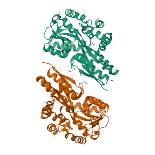
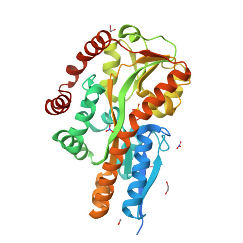
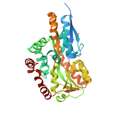
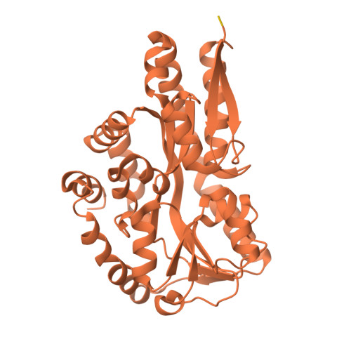
On the function of TRAP substrate-binding proteins: the isethionate-specific binding protein IseP.

Newton-Vesty, M.C.Currie, M.J.Davies, J.S.Panjikar, S.Sethi, A.Whitten, A.E.Tillett, Z.D.Wood, D.M.Wright, J.D.Love, M.J.Allison, T.M.Jamieson, S.A.Mace, P.D.North, R.A.Dobson, R.C.J.

(2024) Biochem J 481: 1901-1920

  • DOI: https://doi.org/10.1042/BCJ20240540
  • Primary Citation of Related Structures:  
    8T9T, 8TE9, 8TQN, 8TRP

  • PubMed Abstract: 

    Bacteria evolve mechanisms to compete for limited resources and survive in new niches. Here we study the mechanism of isethionate import from the sulfate-reducing bacterium Oleidesulfovibrio alaskensis. The catabolism of isethionate by Desulfovibrio species has been implicated in human disease, due to hydrogen sulfide production, and has potential for industrial applications. O. alaskensis employs a tripartite ATP-independent periplasmic (TRAP) transporter (OaIsePQM) to import isethionate, which relies on the substrate-binding protein (OaIseP) to scavenge isethionate and deliver it to the membrane transporter component (OaIseQM) for import into the cell. We determined the binding affinity of isethionate to OaIseP by isothermal titration calorimetry, KD = 0.95 µM (68% CI = 0.6-1.4 µM), which is weaker compared with other TRAP substrate-binding proteins. The X-ray crystal structures of OaIseP in the ligand-free and isethionate-bound forms were obtained and showed that in the presence of isethionate, OaIseP adopts a closed conformation whereby two domains of the protein fold over the substrate. We serendipitously discovered two crystal forms with sulfonate-containing buffers (HEPES and MES) bound in the isethionate-binding site. However, these do not evoke domain closure, presumably because of the larger ligand size. Together, our data elucidate the molecular details of how a TRAP substrate-binding protein binds a sulfonate-containing substrate, rather than a typical carboxylate-containing substrate. These results may inform future antibiotic development to target TRAP transporters and provide insights into protein engineering of TRAP transporter substrate-binding proteins.


  • Organizational Affiliation

    Biomolecular Interaction Centre, School of Biological Sciences, MacDiarmid Institute for Advanced Materials and Nanotechnology, University of Canterbury, Christchurch 8140, New Zealand.


Macromolecules
Find similar proteins by:  (by identity cutoff)  |  3D Structure
Entity ID: 1
MoleculeChains Sequence LengthOrganismDetailsImage
Isethionate-binding periplasmic protein DctP
A, B
336Oleidesulfovibrio alaskensis G20Mutation(s): 0 
Gene Names: dctPDde_1275
UniProt
Find proteins for Q312S0 (Oleidesulfovibrio alaskensis (strain ATCC BAA-1058 / DSM 17464 / G20))
Explore Q312S0 
Go to UniProtKB:  Q312S0
Entity Groups  
Sequence Clusters30% Identity50% Identity70% Identity90% Identity95% Identity100% Identity
UniProt GroupQ312S0
Sequence Annotations
Expand
  • Reference Sequence
Small Molecules
Ligands 3 Unique
IDChains Name / Formula / InChI Key2D Diagram3D Interactions
8X3 (Subject of Investigation/LOI)
Query on 8X3

Download Ideal Coordinates CCD File 
C [auth A],
J [auth B]
2-hydroxyethylsulfonic acid
C2 H6 O4 S
SUMDYPCJJOFFON-UHFFFAOYSA-N
EDO
Query on EDO

Download Ideal Coordinates CCD File 
E [auth A],
F [auth A],
G [auth A],
H [auth A],
K [auth B]
1,2-ETHANEDIOL
C2 H6 O2
LYCAIKOWRPUZTN-UHFFFAOYSA-N
NO3
Query on NO3

Download Ideal Coordinates CCD File 
D [auth A],
I [auth A],
L [auth B],
M [auth B]
NITRATE ION
N O3
NHNBFGGVMKEFGY-UHFFFAOYSA-N
Experimental Data & Validation

Experimental Data

  • Method: X-RAY DIFFRACTION
  • Resolution: 1.25 Å
  • R-Value Free:  0.169 (Depositor), 0.170 (DCC) 
  • R-Value Work:  0.146 (Depositor), 0.147 (DCC) 
Space Group: P 21 21 21
Unit Cell:
Length ( Å )Angle ( ˚ )
a = 72.2α = 90
b = 83.21β = 90
c = 91.91γ = 90
Software Package:
Software NamePurpose
REFMACrefinement
REFMACrefinement
MOLREPphasing
XDSdata reduction
Aimlessdata scaling

Structure Validation

View Full Validation Report


Ligand Structure Quality Assessment 

Created with Raphaël 2.3.0Worse 01 BetterLigand structure goodness of fit to experimental dataBest fitted 8X3Click on this verticalbar to view details

Entry History & Funding Information

Deposition Data


Funding OrganizationLocationGrant Number
Marsden FundNew Zealand--

Revision History  (Full details and data files)

  • Version 1.0: 2024-07-10
  • Version 2.0: 2024-10-02
    Type: Coordinate replacement
    Reason: Model orientation/position
    Changes: Advisory, Atomic model, Data collection, Refinement description, Structure summary
  • Version 2.1: 2025-01-15
    Changes: Database references, Structure summary