10ID | pdb_000010id

Membrane-bound, reversed VP5* trimer (rotavirus spike protein)


Experimental Data Snapshot

  • Method: ELECTRON MICROSCOPY
  • Resolution: 9.54 Å
  • Aggregation State: CELL 
  • Reconstruction Method: SUBTOMOGRAM AVERAGING 

wwPDB Validation   3D Report Full Report


This is version 1.1 of the entry. See complete history


Literature

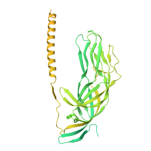
Mechanism of membrane perforation in rotavirus cell entry.

de Sautu, M.Leistner, C.Kirchhausen, T.Jenni, S.Harrison, S.C.

(2026) bioRxiv 

  • DOI: https://doi.org/10.64898/2026.01.21.700916
  • Primary Citation of Related Structures:  
    10IC, 10ID

  • PubMed Abstract: 

    Infectious cell entry by non-enveloped viruses requires delivery of the viral genome -- in many cases enclosed within a large, subviral particle -- across the membrane of an intracellular compartment. Rotaviruses and other double-strand RNA (dsRNA) viruses introduce into their target cells an inner capsid particle, roughly 700 Å in diameter, that does not uncoat further but instead extrudes capped viral mRNA by virtue of RNA-dependent RNA polymerase and capping activities within it. The delivery agent is an outer protein layer of the virion. We describe here use of cryogenic electron tomography (cryo-ET) to visualize the full course of rhesus rotavirus (RRV) entry, from cell attachment and inward budding of the virion to arrival of the subviral particle in the cytosol. The cryo-tomograms and subtomogram averaging of classified subparticles have enabled us to link high-resolution structures of the virion and its components with time series from live-cell fluorescence microscopy and thus to outline the molecular mechanism of each step in the entry process, including the hitherto elusive membrane perforation step needed for transfer of the subviral particle into the cytosol.


  • Organizational Affiliation
    • Department of Biological Chemistry and Molecular Pharmacology, Harvard Medical School, Boston, Massachusetts, United States of America.

Macromolecules
Find similar proteins by:  (by identity cutoff)  |  3D Structure
Entity ID: 1
MoleculeChains Sequence LengthOrganismDetailsImage
Outer capsid protein VP4A [auth 2],
B [auth 3],
C [auth 4]
776Simian rotavirus A strain RRVMutation(s): 0 
UniProt
Find proteins for P12473 (Rotavirus A (strain RVA/Monkey/United States/RRV/1975/G3P5B[3]))
Explore P12473 
Go to UniProtKB:  P12473
Entity Groups  
Sequence Clusters30% Identity50% Identity70% Identity90% Identity95% Identity100% Identity
UniProt GroupP12473
Sequence Annotations
Expand
  • Reference Sequence
Experimental Data & Validation

Experimental Data

  • Method: ELECTRON MICROSCOPY
  • Resolution: 9.54 Å
  • Aggregation State: CELL 
  • Reconstruction Method: SUBTOMOGRAM AVERAGING 
EM Software:
TaskSoftware PackageVersion
RECONSTRUCTIONWarp

Structure Validation

View Full Validation Report


Entry History & Funding Information

Deposition Data


Funding OrganizationLocationGrant Number
National Institutes of Health/National Cancer Institute (NIH/NCI)United StatesR01 CA13202
National Institutes of Health/National Institute of General Medical Sciences (NIH/NIGMS)United StatesR35 GM139386
National Institutes of Health/National Institute Of Allergy and Infectious Diseases (NIH/NIAID)United StatesAI163019

Revision History  (Full details and data files)

  • Version 1.0: 2026-02-04
    Type: Initial release
  • Version 1.1: 2026-03-04
    Changes: Data collection, Database references