1AE9 | pdb_00001ae9

STRUCTURE OF THE LAMBDA INTEGRASE CATALYTIC CORE


Experimental Data Snapshot

  • Method: X-RAY DIFFRACTION
  • Resolution: 1.90 Å
  • R-Value Free: 
    0.232 (Depositor) 
  • R-Value Work: 
    0.199 (Depositor) 
  • R-Value Observed: 
    0.199 (Depositor) 

wwPDB Validation 3D Report Full Report

Validation slider image for 1AE9

This is version 1.4 of the entry. See complete history

Literature

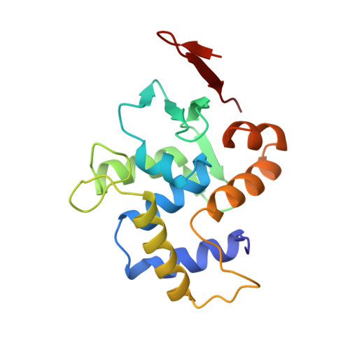
Flexibility in DNA recombination: structure of the lambda integrase catalytic core.

Kwon, H.J.Tirumalai, R.Landy, A.Ellenberger, T.

(1997) Science 276: 126-131

  • DOI: https://doi.org/10.1126/science.276.5309.126
  • Primary Citation Related Structures: 
    1AE9

  • PubMed Abstract: 

    Lambda integrase is archetypic of site-specific recombinases that catalyze intermolecular DNA rearrangements without energetic input. DNA cleavage, strand exchange, and religation steps are linked by a covalent phosphotyrosine intermediate in which Tyr342 is attached to the 3'-phosphate of the DNA cut site. The 1.9 angstrom crystal structure of the integrase catalytic domain reveals a protein fold that is conserved in organisms ranging from archaebacteria to yeast and that suggests a model for interaction with target DNA. The attacking Tyr342 nucleophile is located on a flexible loop about 20 angstroms from a basic groove that contains all the other catalytically essential residues. This bipartite active site can account for several apparently paradoxical features of integrase family recombinases, including the capacity for both cis and trans cleavage of DNA.


  • Organizational Affiliation
    • Department of Biological Chemistry and Molecular Pharmacology, Harvard Medical School, Boston MA 02115, USA.

Macromolecule Content 

  • Total Structure Weight: 40.53 kDa 
  • Atom Count: 2,813 
  • Modeled Residue Count: 337 
  • Deposited Residue Count: 358 
  • Unique protein chains: 1

Macromolecules

Find similar proteins by:|  3D Structure
Entity ID: 1
MoleculeChains  Sequence LengthOrganismDetailsImage
LAMBDA INTEGRASE
A, B
179Lambdavirus lambdaMutation(s): 1 
EC: 3.1 (UniProt), 2.7.7 (UniProt)
UniProt
Find proteins for P03700 (Escherichia phage lambda)
Explore P03700 
Go to UniProtKB:  P03700
Entity Groups
Sequence Clusters30% Identity50% Identity70% Identity90% Identity95% Identity100% Identity
UniProt GroupP03700
Sequence Annotations
Expand
Reference Sequence

Experimental Data & Validation

Experimental Data

  • Method: X-RAY DIFFRACTION
  • Resolution: 1.90 Å
  • R-Value Free:  0.232 (Depositor) 
  • R-Value Work:  0.199 (Depositor) 
  • R-Value Observed: 0.199 (Depositor) 
Space Group: H 3
Unit Cell:
Length ( Å )Angle ( ˚ )
a = 107.32α = 90
b = 107.32β = 90
c = 108.71γ = 120
Software Package:
Software NamePurpose
X-PLORmodel building
X-PLORrefinement
DENZOdata reduction
SCALEPACKdata scaling
X-PLORphasing

Structure Validation

View Full Validation Report


Entry History 

Deposition Data

Revision History  (Full details and data files)

  • Version 1.0: 1997-11-19
    Type: Initial release
  • Version 1.1: 2008-03-24
    Changes: Version format compliance
  • Version 1.2: 2011-07-13
    Changes: Version format compliance
  • Version 1.3: 2021-11-03
    Changes: Database references, Other
  • Version 1.4: 2024-02-07
    Changes: Data collection