1BFR

IRON STORAGE AND ELECTRON TRANSPORT


Experimental Data Snapshot

  • Method: X-RAY DIFFRACTION
  • Resolution: 2.94 Å
  • R-Value Work: 
    0.216 (Depositor) 
  • R-Value Observed: 
    0.216 (Depositor) 

wwPDB Validation   3D Report Full Report


This is version 1.3 of the entry. See complete history


Literature

Structure of a monoclinic crystal from of cyctochrome b1 (Bacterioferritin) from E. coli.

Dautant, A.Meyer, J.B.Yariv, J.Precigoux, G.Sweet, R.M.Kalb, A.J.Frolow, F.

(1998) Acta Crystallogr D Biol Crystallogr 54: 16-24

  • DOI: https://doi.org/10.1107/s0907444997006811
  • Primary Citation of Related Structures:  
    1BFR

  • PubMed Abstract: 

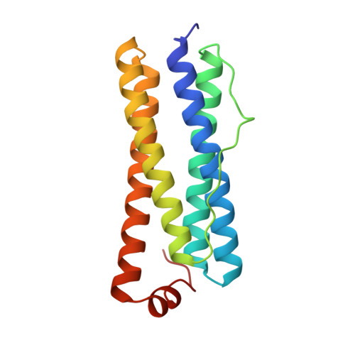
    Crystals of E. coli cytochrome b1, alias bacterioferritin, were grown fr om a low ionic strength solution. The resulting monoclniic P21 structure was solved by molecular replacement and refined using noncrystallographi c symmetries applied to the fundamental unit, consisting of two protein subunits and a single haem. From the Patterson self-rotation results it was shown that the asymmetric unit of the monoclinic crystal consists of 12 such dimers and corresponds to a complete, nearly spherical, molecule of bacterioferritin (M4 = 450 kDa) of 432 point-group symmetry. It is thus the most symmetrical cytochrome. As previously determined for the tetragonal form, the haem is located in a special position on a local twofold axis of the dimer. A bimetal centre is also observed within the four-helix bundle of each monomer; a metal-binding site is located on the fourfold axis.


  • Organizational Affiliation

    Unité Biophysique Structurale, ERS133 CNRS, Université de Bordeaux 1, France.


Macromolecules
Find similar proteins by:  (by identity cutoff)  |  3D Structure
Entity ID: 1
MoleculeChains Sequence LengthOrganismDetailsImage
BACTERIOFERRITIN
A, B, C, D, E
158Escherichia coliMutation(s): 0 
EC: 1.16.3.1
UniProt
Find proteins for P0ABD3 (Escherichia coli (strain K12))
Explore P0ABD3 
Go to UniProtKB:  P0ABD3
Entity Groups  
Sequence Clusters30% Identity50% Identity70% Identity90% Identity95% Identity100% Identity
UniProt GroupP0ABD3
Sequence Annotations
Expand
  • Reference Sequence
Small Molecules
Ligands 2 Unique
IDChains Name / Formula / InChI Key2D Diagram3D Interactions
HEM
Query on HEM

Download Ideal Coordinates CCD File 
AC [auth V]
BB [auth L]
CA [auth B]
FC [auth X]
GB [auth N]
PROTOPORPHYRIN IX CONTAINING FE
C34 H32 Fe N4 O4
KABFMIBPWCXCRK-RGGAHWMASA-L
MN
Query on MN

Download Ideal Coordinates CCD File 
AA [auth B]
AB [auth L]
BA [auth B]
BC [auth W]
CB [auth M]
MANGANESE (II) ION
Mn
WAEMQWOKJMHJLA-UHFFFAOYSA-N
Experimental Data & Validation

Experimental Data

  • Method: X-RAY DIFFRACTION
  • Resolution: 2.94 Å
  • R-Value Work:  0.216 (Depositor) 
  • R-Value Observed: 0.216 (Depositor) 
Space Group: P 1 21 1
Unit Cell:
Length ( Å )Angle ( ˚ )
a = 118.7α = 90
b = 211.6β = 119.1
c = 123.3γ = 90
Software Package:
Software NamePurpose
X-PLORrefinement

Structure Validation

View Full Validation Report


Entry History 

Deposition Data

Revision History  (Full details and data files)

  • Version 1.0: 1996-06-20
    Type: Initial release
  • Version 1.1: 2008-03-24
    Changes: Version format compliance
  • Version 1.2: 2011-07-13
    Changes: Version format compliance
  • Version 1.3: 2024-02-07
    Changes: Data collection, Database references, Derived calculations, Other