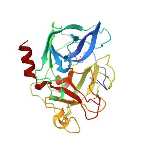
1BTU

PORCINE PANCREATIC ELASTASE COMPLEXED WITH (3S, 4R)-1-TOLUENESULPHONYL-3-ETHYL-AZETIDIN-2-ONE-4-CARBOXYLIC ACID


Experimental Data Snapshot

  • Method: X-RAY DIFFRACTION
  • Resolution: 1.60 Å
  • R-Value Free: 0.220 
  • R-Value Observed: 0.192 

wwPDB Validation   3D Report Full Report


This is version 1.3 of the entry. See complete history


Literature

Inhibition of elastase by N-sulfonylaryl beta-lactams: anatomy of a stable acyl-enzyme complex.

Wilmouth, R.C.Westwood, N.J.Anderson, K.Brownlee, W.Claridge, T.D.Clifton, I.J.Pritchard, G.J.Aplin, R.T.Schofield, C.J.

(1998) Biochemistry 37: 17506-17513

  • DOI: https://doi.org/10.1021/bi9816249
  • Primary Citation of Related Structures:  
    1BTU

  • PubMed Abstract: 

    beta-Lactam inhibitors of transpeptidase enzymes involved in cell wall biosynthesis remain among the most important therapeutic agents in clinical use. beta-Lactams have more recently been developed as inhibitors of serine proteases including elastase. All therapeutically useful beta-lactam inhibitors operate via mechanisms resulting in the formation of hydrolytically stable acyl-enzyme complexes. Presently, it is difficult to predict which beta-lactams will form stable acyl-enzyme complexes with serine enzymes. Further, the factors that result in the seemingly special nature of beta-lactams versus other acylating agents are unclear-if indeed they exist. Here we present the 1.6 A resolution crystal structure of a stable acyl-enzyme complex formed between porcine pancreatic elastase and a representative monocyclic beta-lactam, which forms a simple acyl-enzyme. The structure shows that the ester carbonyl is not located within the oxyanion hole and the "hydrolytic" water is displaced. Combined with additional kinetic and mass spectrometric data, the structure allows the rationalization of the low degree of hydrolytic lability observed for the beta-lactam-derived acyl-enzyme complex.


  • Organizational Affiliation

    The Oxford Centre for Molecular Sciences, The Dyson Perrins Laboratory, Oxford, United Kingdom.


Macromolecules
Find similar proteins by:  (by identity cutoff)  |  3D Structure
Entity ID: 1
MoleculeChains Sequence LengthOrganismDetailsImage
ELASTASE240Sus scrofaMutation(s): 0 
EC: 3.4.21.36
UniProt
Find proteins for P00772 (Sus scrofa)
Explore P00772 
Go to UniProtKB:  P00772
Entity Groups  
Sequence Clusters30% Identity50% Identity70% Identity90% Identity95% Identity100% Identity
UniProt GroupP00772
Sequence Annotations
Expand
  • Reference Sequence
Small Molecules
Ligands 3 Unique
IDChains Name / Formula / InChI Key2D Diagram3D Interactions
2BL
Query on 2BL

Download Ideal Coordinates CCD File 
D [auth A](3R)-3-ethyl-N-[(4-methylphenyl)sulfonyl]-L-aspartic acid
C13 H17 N O6 S
KPHLTCNXHCHMOW-MNOVXSKESA-N
SO4
Query on SO4

Download Ideal Coordinates CCD File 
C [auth A]SULFATE ION
O4 S
QAOWNCQODCNURD-UHFFFAOYSA-L
CA
Query on CA

Download Ideal Coordinates CCD File 
B [auth A]CALCIUM ION
Ca
BHPQYMZQTOCNFJ-UHFFFAOYSA-N
Experimental Data & Validation

Experimental Data

  • Method: X-RAY DIFFRACTION
  • Resolution: 1.60 Å
  • R-Value Free: 0.220 
  • R-Value Observed: 0.192 
  • Space Group: P 21 21 21
Unit Cell:
Length ( Å )Angle ( ˚ )
a = 50.57α = 90
b = 57.93β = 90
c = 74.72γ = 90
Software Package:
Software NamePurpose
DENZOdata reduction
SCALEPACKdata scaling
SHELXL-97model building
X-PLORmodel building
SHELXL-97refinement
X-PLORrefinement
SHELXL-97phasing
X-PLORphasing

Structure Validation

View Full Validation Report


Entry History 

Deposition Data

Revision History  (Full details and data files)

  • Version 1.0: 1999-02-16
    Type: Initial release
  • Version 1.1: 2008-03-24
    Changes: Version format compliance
  • Version 1.2: 2011-07-13
    Changes: Non-polymer description, Version format compliance
  • Version 1.3: 2024-10-16
    Changes: Data collection, Database references, Derived calculations, Structure summary