1CQP

CRYSTAL STRUCTURE ANALYSIS OF THE COMPLEX LFA-1 (CD11A) I-DOMAIN / LOVASTATIN AT 2.6 A RESOLUTION


Experimental Data Snapshot

  • Method: X-RAY DIFFRACTION
  • Resolution: 2.60 Å
  • R-Value Free: 
    0.257 (Depositor), 0.180 (DCC) 
  • R-Value Work: 
    0.190 (Depositor), 0.170 (DCC) 
  • R-Value Observed: 
    0.190 (Depositor) 

wwPDB Validation   3D Report Full Report


Ligand Structure Quality Assessment 

Created with Raphaël 2.3.0Worse 01 BetterLigand structure goodness of fit to experimental dataBest fitted 803Click on this verticalbar to view details

This is version 1.4 of the entry. See complete history


Literature

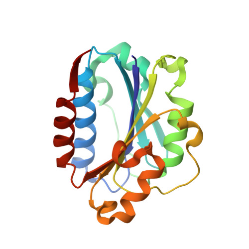
Structural basis for LFA-1 inhibition upon lovastatin binding to the CD11a I-domain.

Kallen, J.Welzenbach, K.Ramage, P.Geyl, D.Kriwacki, R.Legge, G.Cottens, S.Weitz-Schmidt, G.Hommel, U.

(1999) J Mol Biol 292: 1-9

  • DOI: https://doi.org/10.1006/jmbi.1999.3047
  • Primary Citation of Related Structures:  
    1CQP

  • PubMed Abstract: 

    The lymphocyte function-associated antigen (LFA-1) belongs to the family of beta2-integrins and plays an important role in T-cell activation and leukocyte migration to sites of inflammation. We report here that lovastatin, a drug clinically used for lowering cholesterol levels, inhibits the interaction of human LFA-1 with its counter-receptor intercellular adhesion molecule-1. Using nuclear magnetic resonance spectroscopy and X-ray crystallography we show that the inhibitor binds to a highly conserved domain of the LFA-1 alpha-chain called the I-domain. The first three-dimensional structure of an integrin inhibitor bound to its receptor reveals atomic details for a hitherto unknown mode of LFA-1 inhibition. It also sheds light into possible mechanisms of LFA-1 mediated signalling and will support the design of novel anti-adhesive and immunosuppressive drugs.


  • Organizational Affiliation

    Preclinical Research, NOVARTIS PHARMA AG, Basel, CH 4002, Switzerland.


Macromolecules
Find similar proteins by:  (by identity cutoff)  |  3D Structure
Entity ID: 1
MoleculeChains Sequence LengthOrganismDetailsImage
ANTIGEN CD11A (P180)
A, B
182Homo sapiensMutation(s): 0 
UniProt & NIH Common Fund Data Resources
Find proteins for P20701 (Homo sapiens)
Explore P20701 
Go to UniProtKB:  P20701
PHAROS:  P20701
GTEx:  ENSG00000005844 
Entity Groups  
Sequence Clusters30% Identity50% Identity70% Identity90% Identity95% Identity100% Identity
UniProt GroupP20701
Sequence Annotations
Expand
  • Reference Sequence
Experimental Data & Validation

Experimental Data

  • Method: X-RAY DIFFRACTION
  • Resolution: 2.60 Å
  • R-Value Free:  0.257 (Depositor), 0.180 (DCC) 
  • R-Value Work:  0.190 (Depositor), 0.170 (DCC) 
  • R-Value Observed: 0.190 (Depositor) 
Space Group: P 21 21 21
Unit Cell:
Length ( Å )Angle ( ˚ )
a = 72.7α = 90
b = 77.7β = 90
c = 91.8γ = 90
Software Package:
Software NamePurpose
X-PLORmodel building
X-PLORrefinement
DENZOdata reduction
SCALEPACKdata scaling
X-PLORphasing

Structure Validation

View Full Validation Report


Ligand Structure Quality Assessment 

Created with Raphaël 2.3.0Worse 01 BetterLigand structure goodness of fit to experimental dataBest fitted 803Click on this verticalbar to view details

Entry History 

Deposition Data

Revision History  (Full details and data files)

  • Version 1.0: 2000-08-07
    Type: Initial release
  • Version 1.1: 2008-04-27
    Changes: Version format compliance
  • Version 1.2: 2011-07-13
    Changes: Version format compliance
  • Version 1.3: 2014-11-12
    Changes: Structure summary
  • Version 1.4: 2024-02-07
    Changes: Data collection, Database references, Derived calculations