1DUB

2-ENOYL-COA HYDRATASE, DATA COLLECTED AT 100 K, PH 6.5


Experimental Data Snapshot

  • Method: X-RAY DIFFRACTION
  • Resolution: 2.50 Å
  • R-Value Free: 
    0.265 (Depositor) 
  • R-Value Work: 
    0.209 (Depositor), 0.190 (DCC) 
  • R-Value Observed: 
    0.209 (Depositor) 

wwPDB Validation   3D Report Full Report


Ligand Structure Quality Assessment 

Created with Raphaël 2.3.0Worse 01 BetterLigand structure goodness of fit to experimental dataBest fitted CAAClick on this verticalbar to view details

This is version 1.3 of the entry. See complete history


Literature

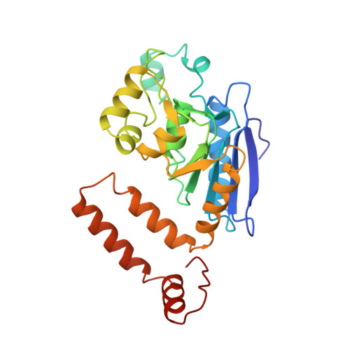
Crystal structure of enoyl-coenzyme A (CoA) hydratase at 2.5 angstroms resolution: a spiral fold defines the CoA-binding pocket.

Engel, C.K.Mathieu, M.Zeelen, J.P.Hiltunen, J.K.Wierenga, R.K.

(1996) EMBO J 15: 5135-5145

  • Primary Citation of Related Structures:  
    1DUB

  • PubMed Abstract: 

    The crystal structure of rat liver mitochondrial enoyl-coenzyme A (CoA) hydratase complexed with the potent inhibitor acetoacetyl-CoA has been refined at 2.5 angstroms resolution. This enzyme catalyses the reversible addition of water to alpha,beta-unsaturated enoyl-CoA thioesters, with nearly diffusion-controlled reaction rates for the best substrates. Enoyl-CoA hydratase is a hexamer of six identical subunits of 161 kDa molecular mass for the complex. The hexamer is a dimer of trimers. The monomer is folded into a right-handed spiral of four turns, followed by two small domains which are involved in trimerization. Each turn of the spiral consists of two beta-strands and an alpha-helix. The mechanism for the hydratase/dehydratase reaction follows a syn-stereochemistry, a preference that is opposite to the nonenzymatic reaction. The active-site architecture agrees with this stereochemistry. It confirms the importance of Glu164 as the catalytic acid for providing the alpha-proton during the hydratase reaction. It also shows the importance of Glu144 as the catalytic base for the activation of a water molecule in the hydratase reaction. The comparison of an unliganded and a liganded active site within the same crystal form shows a water molecule in the unliganded subunit. This water molecule is bound between the two catalytic glutamates and could serve as the activated water during catalysis.


  • Organizational Affiliation

    European Molecular Biology Laboratory, Heidelberg, Germany.


Macromolecules
Find similar proteins by:  (by identity cutoff)  |  3D Structure
Entity ID: 1
MoleculeChains Sequence LengthOrganismDetailsImage
2-ENOYL-COA HYDRATASE
A, B, C, D, E
261Rattus norvegicusMutation(s): 0 
EC: 4.2.1.17 (PDB Primary Data), 5.3.3.8 (UniProt)
UniProt
Find proteins for P14604 (Rattus norvegicus)
Explore P14604 
Go to UniProtKB:  P14604
Entity Groups  
Sequence Clusters30% Identity50% Identity70% Identity90% Identity95% Identity100% Identity
UniProt GroupP14604
Sequence Annotations
Expand
  • Reference Sequence
Experimental Data & Validation

Experimental Data

  • Method: X-RAY DIFFRACTION
  • Resolution: 2.50 Å
  • R-Value Free:  0.265 (Depositor) 
  • R-Value Work:  0.209 (Depositor), 0.190 (DCC) 
  • R-Value Observed: 0.209 (Depositor) 
Space Group: P 21 21 21
Unit Cell:
Length ( Å )Angle ( ˚ )
a = 75.972α = 90
b = 93.635β = 90
c = 246.818γ = 90
Software Package:
Software NamePurpose
MLPHAREphasing
X-PLORrefinement
DENZOdata reduction

Structure Validation

View Full Validation Report


Ligand Structure Quality Assessment 

Created with Raphaël 2.3.0Worse 01 BetterLigand structure goodness of fit to experimental dataBest fitted CAAClick on this verticalbar to view details

Entry History 

Deposition Data

Revision History  (Full details and data files)

  • Version 1.0: 1997-07-07
    Type: Initial release
  • Version 1.1: 2008-03-24
    Changes: Version format compliance
  • Version 1.2: 2011-07-13
    Changes: Version format compliance
  • Version 1.3: 2024-02-07
    Changes: Data collection, Database references, Derived calculations