1ERJ

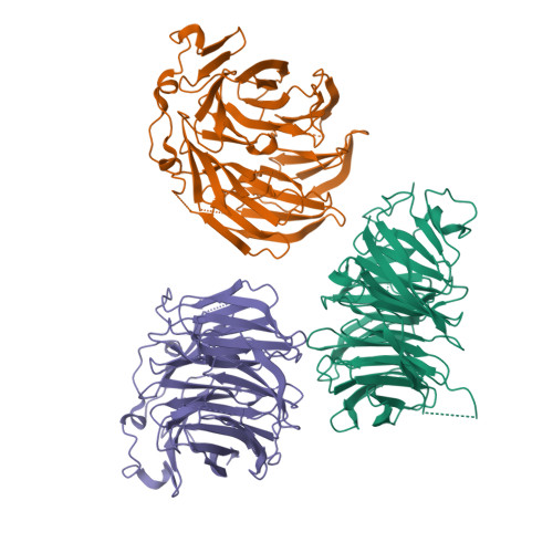
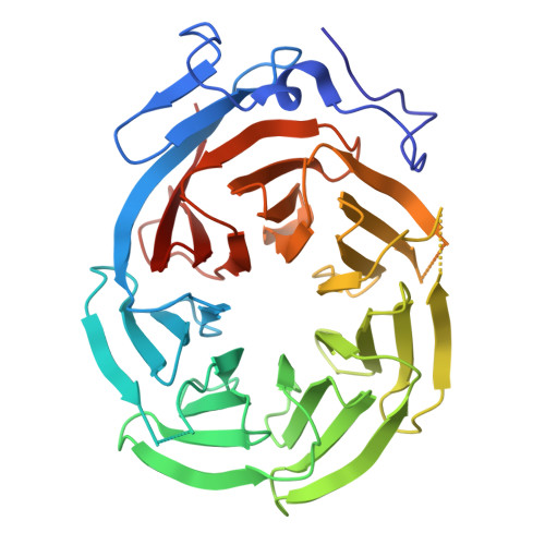
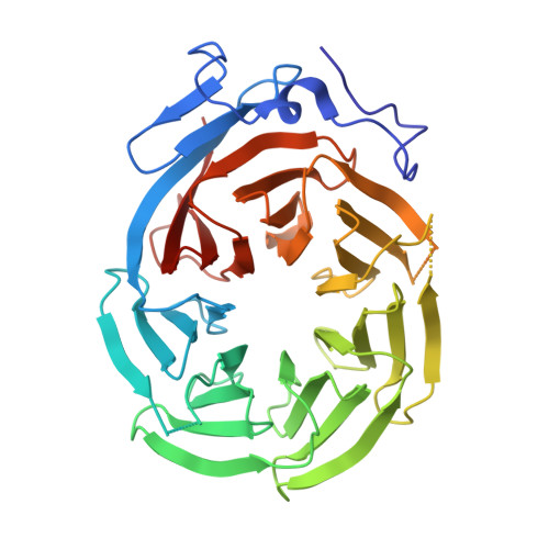
CRYSTAL STRUCTURE OF THE C-TERMINAL WD40 DOMAIN OF TUP1


Experimental Data Snapshot

  • Method: X-RAY DIFFRACTION
  • Resolution: 2.30 Å
  • R-Value Free: 0.266 
  • R-Value Work: 0.228 

wwPDB Validation   3D Report Full Report


This is version 1.5 of the entry. See complete history


Literature

Structure of the C-terminal domain of Tup1, a corepressor of transcription in yeast.

Sprague, E.R.Redd, M.J.Johnson, A.D.Wolberger, C.

(2000) EMBO J 19: 3016-3027

  • DOI: https://doi.org/10.1093/emboj/19.12.3016
  • Primary Citation of Related Structures:  
    1ERJ

  • PubMed Abstract: 

    The Tup1-Ssn6 corepressor complex regulates the expression of several sets of genes, including genes that specify mating type in the yeast Saccharomyces cerevisiae. Repression of mating-type genes occurs when Tup1-Ssn6 is brought to the DNA by the Matalpha2 DNA-binding protein and assembled upstream of a- and haploid-specific genes. We have determined the 2.3 A X-ray crystal structure of the C-terminal domain of Tup1 (accesion No. 1ERJ), a 43 kDa fragment that contains seven copies of the WD40 sequence motif and binds to the Matalpha2 protein. Moreover, this portion of the protein can partially substitute for full-length Tup1 in bringing about transcriptional repression. The structure reveals a seven-bladed beta propeller with an N-terminal subdomain that is anchored to the side of the propeller and extends the beta sheet of one of the blades. Point mutations in Tup1 that specifically affect the Tup1-Matalpha2 interaction cluster on one surface of the propeller. We identified regions of Tup1 that are conserved among the fungal Tup1 homologs and may be important in protein-protein interactions with additional components of the Tup1-mediated repression pathways.


  • Organizational Affiliation

    Department of Biophysics and Biophysical Chemistry and Howard Hughes Medical Institute, Johns Hopkins University School of Medicine, Baltimore, MD 21205, USA.


Macromolecules
Find similar proteins by:  (by identity cutoff)  |  3D Structure
Entity ID: 1
MoleculeChains Sequence LengthOrganismDetailsImage
TRANSCRIPTIONAL REPRESSOR TUP1
A, B, C
393Saccharomyces cerevisiaeMutation(s): 0 
UniProt
Find proteins for P16649 (Saccharomyces cerevisiae (strain ATCC 204508 / S288c))
Explore P16649 
Go to UniProtKB:  P16649
Entity Groups  
Sequence Clusters30% Identity50% Identity70% Identity90% Identity95% Identity100% Identity
UniProt GroupP16649
Sequence Annotations
Expand
  • Reference Sequence
Experimental Data & Validation

Experimental Data

  • Method: X-RAY DIFFRACTION
  • Resolution: 2.30 Å
  • R-Value Free: 0.266 
  • R-Value Work: 0.228 
  • Space Group: P 31
Unit Cell:
Length ( Å )Angle ( ˚ )
a = 119.28α = 90
b = 119.28β = 90
c = 77.07γ = 120
Software Package:
Software NamePurpose
MLPHAREphasing
CNSrefinement
DENZOdata reduction
SCALEPACKdata scaling

Structure Validation

View Full Validation Report


Entry History 

Deposition Data

Revision History  (Full details and data files)

  • Version 1.0: 2000-07-26
    Type: Initial release
  • Version 1.1: 2008-04-27
    Changes: Version format compliance
  • Version 1.2: 2011-07-13
    Changes: Version format compliance
  • Version 1.3: 2017-08-09
    Changes: Refinement description, Source and taxonomy
  • Version 1.4: 2022-12-21
    Changes: Database references
  • Version 1.5: 2024-05-22
    Changes: Data collection