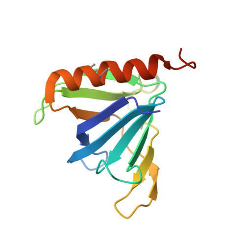
1FHX

Structure of the pleckstrin homology domain from GRP1 in complex with inositol 1,3,4,5-tetrakisphosphate


Experimental Data Snapshot

  • Method: X-RAY DIFFRACTION
  • Resolution: 2.50 Å
  • R-Value Free: 0.286 
  • R-Value Work: 0.228 
  • R-Value Observed: 0.228 

Starting Model: experimental
View more details

wwPDB Validation   3D Report Full Report


This is version 1.5 of the entry. See complete history


Literature

Structural basis for discrimination of 3-phosphoinositides by pleckstrin homology domains

Ferguson, K.M.Kavran, J.M.Sankaran, V.G.Fournier, E.Isakoff, S.J.Skolnik, E.Y.Lemmon, M.A.

(2000) Mol Cell 6: 373-384

  • DOI: https://doi.org/10.1016/s1097-2765(00)00037-x
  • Primary Citation of Related Structures:  
    1FAO, 1FB8, 1FHW, 1FHX

  • PubMed Abstract: 

    Pleckstrin homology (PH) domains are protein modules of around 120 amino acids found in many proteins involved in cellular signaling. Certain PH domains drive signal-dependent membrane recruitment of their host proteins by binding strongly and specifically to lipid second messengers produced by agonist-stimulated phosphoinositide 3-kinases (PI 3-Ks). We describe X-ray crystal structures of two different PH domains bound to Ins(1,3,4,5)P4, the head group of the major PI 3-K product PtdIns(3,4,5)P3. One of these PH domains (from Grp1) is PtdIns(3,4,5)P3 specific, while the other (from DAPP1/PHISH) binds strongly to both PtdIns(3,4,5)P3 and its 5'-dephosphorylation product, PtdIns(3,4)P2. Comparison of the two structures provides an explanation for the distinct phosphoinositide specificities of the two PH domains and allows us to predict the 3-phosphoinositide selectivity of uncharacterized PH domains.


  • Organizational Affiliation

    Department of Biochemistry and Biophysics and The Johnson Foundation, University of Pennsylvania School of Medicine, Philadelphia 19104, USA.


Macromolecules
Find similar proteins by:  (by identity cutoff)  |  3D Structure
Entity ID: 1
MoleculeChains Sequence LengthOrganismDetailsImage
GUANINE NUCLEOTIDE EXCHANGE FACTOR AND INTEGRIN BINDING PROTEIN HOMOLOG GRP1
A, B
129Mus musculusMutation(s): 2 
Membrane Entity: Yes 
UniProt
Find proteins for O08967 (Mus musculus)
Explore O08967 
Go to UniProtKB:  O08967
Entity Groups  
Sequence Clusters30% Identity50% Identity70% Identity90% Identity95% Identity100% Identity
UniProt GroupO08967
Sequence Annotations
Expand
  • Reference Sequence
Small Molecules
Modified Residues  1 Unique
IDChains TypeFormula2D DiagramParent
MSE
Query on MSE
A, B
L-PEPTIDE LINKINGC5 H11 N O2 SeMET
Binding Affinity Annotations 
IDSourceBinding Affinity
4IP BindingDB:  1FHX Kd: 100 (nM) from 1 assay(s)
Experimental Data & Validation

Experimental Data

  • Method: X-RAY DIFFRACTION
  • Resolution: 2.50 Å
  • R-Value Free: 0.286 
  • R-Value Work: 0.228 
  • R-Value Observed: 0.228 
  • Space Group: P 21 21 21
Unit Cell:
Length ( Å )Angle ( ˚ )
a = 56.09α = 90
b = 64.54β = 90
c = 69.73γ = 90
Software Package:
Software NamePurpose
DENZOdata reduction
SCALEPACKdata scaling
AMoREphasing
CNSrefinement

Structure Validation

View Full Validation Report


Entry History 

Deposition Data

Revision History  (Full details and data files)

  • Version 1.0: 2000-08-23
    Type: Initial release
  • Version 1.1: 2008-04-27
    Changes: Version format compliance
  • Version 1.2: 2011-07-13
    Changes: Derived calculations, Version format compliance
  • Version 1.3: 2023-08-09
    Changes: Data collection, Database references, Derived calculations, Refinement description
  • Version 1.4: 2023-11-15
    Changes: Data collection
  • Version 1.5: 2024-10-09
    Changes: Structure summary