Crystallographic studies of the catalytic and a second site in fumarase C from Escherichia coli.
Weaver, T., Banaszak, L.(1996) Biochemistry 35: 13955-13965
- PubMed: 8909293 Search on PubMed
- DOI: https://doi.org/10.1021/bi9614702
- Primary Citation Related Structures:
1FUO, 1FUP, 1FUQ - PubMed Abstract:
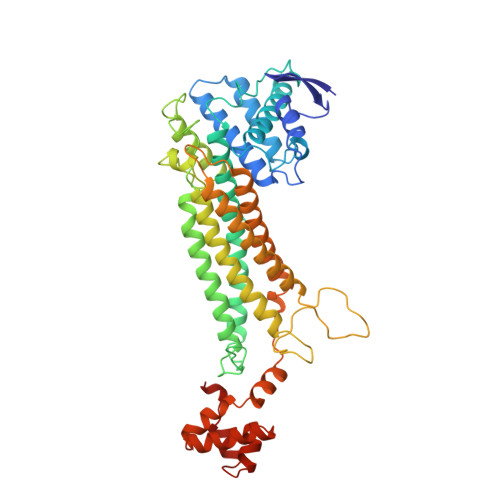
Fumarase C catalyzes the stereospecific interconversion of fumarate to L-malate as part of the metabolic citric acid or Kreb's cycle. The recent three-dimensional structure of fumarase C from Escherichia coli has identified a binding site for anions which is generated by side chains from three of the four subunits within the tetramer (Weaver et al., 1995). These same side chains are found in the three most highly conserved regions within the class II fumarase superfamily. The site was initially characterized by crystallographic studies through the binding of a heavy atom derivative, tungstate. A number of additional crystallographic structures using fumarase crystals with bound inhibitors and poor substrates have now been studied. The new structures have both confirmed the originally proposed active site, site A, and led to the discovery of a novel second binding site that is structurally nearby, site B. Site A utilizes a combination of residues, including H188, T187, K324, N326, T100, N141, S139, and S140, to form direct hydrogen bonds to each of the inhibitors. The A-site has been demonstrated by studying crystalline fumarase with the bound competitive inhibitors-citrate and 1,2,4,5-benzenetetracarboxylic acid. The crystal structure of fumarase C with beta-(trimethylsilyl)maleate, a cis substrate for fumarase, has led to the discovery of the second site or B-site. Sites A and B have different properties in terms of their three-dimensional structures. Site B, for example, is formed by atoms from only one of the subunits within the tetramer and mainly by atoms from a pi-helix between residues H129 through N135. The crystal structures show that the two locations are separated by approximately 12 A. A highly coordinated buried water molecule is also found at the active or A-site. The high-resolution crystal structures describe both sites, and atoms near the A-site are used to propose a likely enzyme/substrate complex.
- Department of Biochemistry, University of Minnesota, Minneapolis 55455, USA.
Organizational Affiliation: