1GWS

hexadecaheme high molecular weight cytochrome Hmc from Desulfovibrio vulgaris Hildenborough


Experimental Data Snapshot

  • Method: X-RAY DIFFRACTION
  • Resolution: 2.40 Å
  • R-Value Free: 
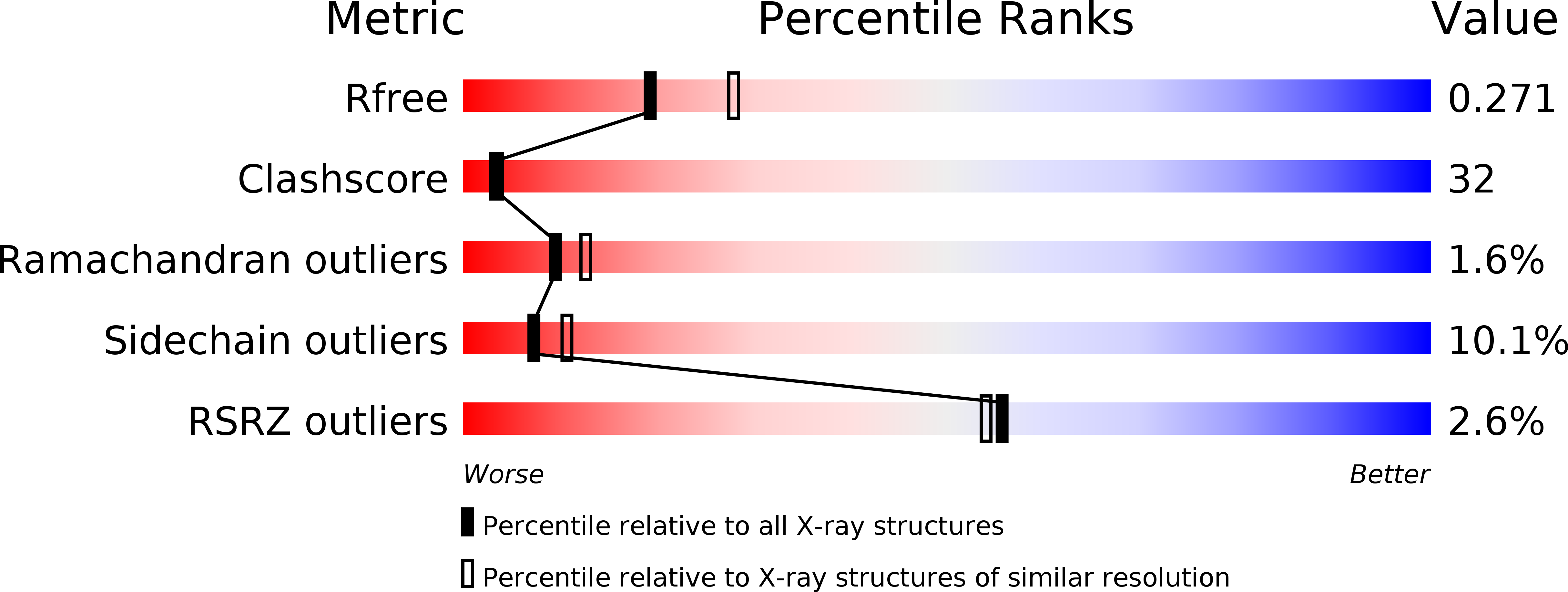
    0.276 (Depositor), 0.271 (DCC) 
  • R-Value Work: 
    0.201 (Depositor), 0.199 (DCC) 

wwPDB Validation   3D Report Full Report


Ligand Structure Quality Assessment 

Created with Raphaël 2.3.0Worse 01 BetterLigand structure goodness of fit to experimental dataBest fitted HECClick on this verticalbar to view details

This is version 2.1 of the entry. See complete history


Literature

The Crystal Structure of the Hexadaca-Heme Cytochrome Hmc and a Structural Model of its Complex with Cytochrome C3

Czjzek, M.Elantak, L.Zamboni, V.Morelli, X.Dolla, A.Guerlesquin, F.Bruschi, M.

(2002) Structure 10: 1677

  • DOI: https://doi.org/10.1016/s0969-2126(02)00909-7
  • Primary Citation of Related Structures:  
    1GWS

  • PubMed Abstract: 

    Sulfate-reducing bacteria contain a variety of multi-heme c-type cytochromes. The cytochrome of highest molecular weight (Hmc) contains 16 heme groups and is part of a transmembrane complex involved in the sulfate respiration pathway. We present the 2.42 A resolution crystal structure of the Desulfovibrio vulgaris Hildenborough cytochrome Hmc and a structural model of the complex with its physiological electron transfer partner, cytochrome c(3), obtained by NMR restrained soft-docking calculations. The Hmc is composed of three domains, which exist independently in different sulfate-reducing species, namely cytochrome c(3), cytochrome c(7), and Hcc. The complex involves the last heme at the C-terminal region of the V-shaped Hmc and heme 4 of cytochrome c(3), and represents an example for specific cytochrome-cytochrome interaction.


  • Organizational Affiliation

    Architecture et Fonction des Macromolécules Biologiques, IBSM-CNRS et Université Aix-Marseille I et II, 31 Chemin Joseph-Aiguier, 13402 Marseille cedex 20, France. czjzek@afmb.cnrs-mrs.fr


Macromolecules
Find similar proteins by:  (by identity cutoff)  |  3D Structure
Entity ID: 1
MoleculeChains Sequence LengthOrganismDetailsImage
HIGH-MOLECULAR-WEIGHT CYTOCHROME C545Nitratidesulfovibrio vulgaris str. HildenboroughMutation(s): 0 
UniProt
Find proteins for P24092 (Nitratidesulfovibrio vulgaris (strain ATCC 29579 / DSM 644 / CCUG 34227 / NCIMB 8303 / VKM B-1760 / Hildenborough))
Explore P24092 
Go to UniProtKB:  P24092
Entity Groups  
Sequence Clusters30% Identity50% Identity70% Identity90% Identity95% Identity100% Identity
UniProt GroupP24092
Sequence Annotations
Expand
  • Reference Sequence
Small Molecules
Ligands 1 Unique
IDChains Name / Formula / InChI Key2D Diagram3D Interactions
HEC
Query on HEC

Download Ideal Coordinates CCD File 
B [auth A]
C [auth A]
D [auth A]
E [auth A]
F [auth A]
HEME C
C34 H34 Fe N4 O4
HXQIYSLZKNYNMH-LJNAALQVSA-N
Experimental Data & Validation

Experimental Data

  • Method: X-RAY DIFFRACTION
  • Resolution: 2.40 Å
  • R-Value Free:  0.276 (Depositor), 0.271 (DCC) 
  • R-Value Work:  0.201 (Depositor), 0.199 (DCC) 
Space Group: P 62
Unit Cell:
Length ( Å )Angle ( ˚ )
a = 108.386α = 90
b = 108.386β = 90
c = 102.809γ = 120
Software Package:
Software NamePurpose
REFMACrefinement
DENZOdata reduction
SCALAdata scaling
SHARPphasing

Structure Validation

View Full Validation Report


Ligand Structure Quality Assessment 

Created with Raphaël 2.3.0Worse 01 BetterLigand structure goodness of fit to experimental dataBest fitted HECClick on this verticalbar to view details

Entry History 

Deposition Data

Revision History  (Full details and data files)

  • Version 1.0: 2003-02-13
    Type: Initial release
  • Version 1.1: 2011-07-13
    Changes: Advisory, Version format compliance
  • Version 2.0: 2019-09-25
    Changes: Advisory, Atomic model, Data collection, Derived calculations, Experimental preparation, Non-polymer description, Other, Structure summary
  • Version 2.1: 2024-10-23
    Changes: Data collection, Database references, Derived calculations, Structure summary