1HAC | pdb_00001hac

CROSSLINKED HAEMOGLOBIN


Experimental Data Snapshot

  • Method: X-RAY DIFFRACTION
  • Resolution: 2.60 Å
  • R-Value Work: 
    0.154 (Depositor) 

wwPDB Validation 3D Report Full Report

Validation slider image for 1HAC

This is version 1.3 of the entry. See complete history

Literature

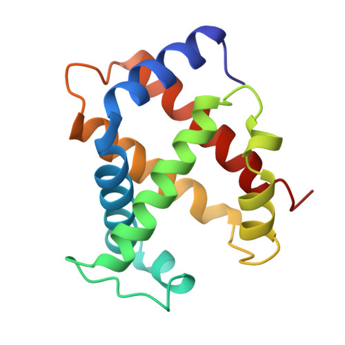
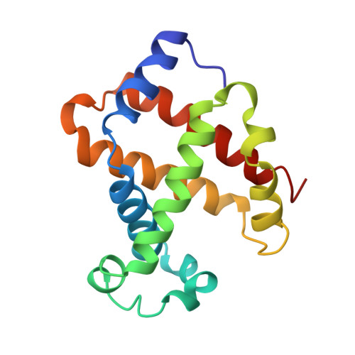
Allosteric intermediates indicate R2 is the liganded hemoglobin end state.

Schumacher, M.A.Zheleznova, E.E.Poundstone, K.S.Kluger, R.Jones, R.T.Brennan, R.G.

(1997) Proc Natl Acad Sci U S A 94: 7841-7844

  • DOI: https://doi.org/10.1073/pnas.94.15.7841
  • Primary Citation Related Structures: 
    1HAB, 1HAC

  • PubMed Abstract: 

    Hemoglobin has been a long-standing paradigm for understanding protein allostery. Here, the x-ray structures of two chemically crosslinked, fully liganded hemoglobins, alpha2beta82CA82beta and alpha2beta82ND82beta, are described at 2.3 A and 2.6 A resolution, respectively. Strikingly, these crosslinked hemoglobins assume intermediate conformations that lie between those of R and the controversial liganded hemoglobin state R2 rather than between R and T. Thus, these structures support only a T left and right arrow R left and right arrow R2 allosteric pathway and underscore the physiological importance of the R2 conformation.


  • Organizational Affiliation
    • Department of Biochemistry and Molecular Biology, Oregon Health Sciences University, 3181 SW Sam Jackson Park Road, Portland, OR 97201-3098, USA.

Macromolecule Content 

  • Total Structure Weight: 64.88 kDa 
  • Atom Count: 4,623 
  • Modeled Residue Count: 574 
  • Deposited Residue Count: 574 
  • Unique protein chains: 2

Macromolecules

Find similar proteins by:|  3D Structure
Entity ID: 1
MoleculeChains  Sequence LengthOrganismDetailsImage
HEMOGLOBIN A
A, C
141Homo sapiensMutation(s): 0 
UniProt & NIH Common Fund Data Resources
Find proteins for P69905 (Homo sapiens)
Explore P69905 
Go to UniProtKB:  P69905
Entity Groups
Sequence Clusters30% Identity50% Identity70% Identity90% Identity95% Identity100% Identity
UniProt GroupP69905
Sequence Annotations
Expand
Reference Sequence
Find similar proteins by:|  3D Structure
Entity ID: 2
MoleculeChains  Sequence LengthOrganismDetailsImage
HEMOGLOBIN A
B, D
146Homo sapiensMutation(s): 0 
UniProt & NIH Common Fund Data Resources
Find proteins for P68871 (Homo sapiens)
Explore P68871 
Go to UniProtKB:  P68871
PHAROS:  P68871
GTEx:  ENSG00000244734 
Entity Groups
Sequence Clusters30% Identity50% Identity70% Identity90% Identity95% Identity100% Identity
UniProt GroupP68871
Sequence Annotations
Expand
Reference Sequence

Small Molecules

Ligands 3 Unique
IDChains Name / Formula / InChI Key2D Diagram3D Interactions
HEM

Query on HEM


Download:Ideal Coordinates CCD File
E [auth A],
G [auth B],
J [auth C],
L [auth D]
PROTOPORPHYRIN IX CONTAINING FE
C34 H32 Fe N4 O4
KABFMIBPWCXCRK-RGGAHWMASA-L
NDD

Query on NDD


Download:Ideal Coordinates CCD File
I [auth B]2,6-DICARBOXYNAPHTHALENE
C12 H8 O4
RXOHFPCZGPKIRD-UHFFFAOYSA-N
CMO

Query on CMO


Download:Ideal Coordinates CCD File
F [auth A],
H [auth B],
K [auth C],
M [auth D]
CARBON MONOXIDE
C O
UGFAIRIUMAVXCW-UHFFFAOYSA-N

Experimental Data & Validation

Experimental Data

  • Method: X-RAY DIFFRACTION
  • Resolution: 2.60 Å
  • R-Value Work:  0.154 (Depositor) 
Space Group: P 21 21 21
Unit Cell:
Length ( Å )Angle ( ˚ )
a = 86.84α = 90
b = 87.11β = 90
c = 97.63γ = 90
Software Package:
Software NamePurpose
TNTrefinement
ADSCdata collection

Structure Validation

View Full Validation Report


Entry History 

Deposition Data

Revision History  (Full details and data files)

  • Version 1.0: 1997-11-12
    Type: Initial release
  • Version 1.1: 2008-03-03
    Changes: Version format compliance
  • Version 1.2: 2011-07-13
    Changes: Version format compliance
  • Version 1.3: 2024-11-20
    Changes: Data collection, Database references, Derived calculations, Structure summary