1HM5 | pdb_00001hm5

CRYSTAL STRUCTURE ANALYSIS OF THE RABBIT D-GLUCOSE 6-PHOSPHATE ISOMERASE (NO LIGAND BOUND)


Experimental Data Snapshot

  • Method: X-RAY DIFFRACTION
  • Resolution: 1.80 Å
  • R-Value Free: 
    0.212 (Depositor), 0.212 (DCC) 
  • R-Value Work: 
    0.183 (Depositor), 0.183 (DCC) 
  • R-Value Observed: 
    0.183 (Depositor) 

Starting Model: experimental
View more details

wwPDB Validation 3D Report Full Report

Validation slider image for 1HM5

This is version 1.4 of the entry. See complete history

Literature

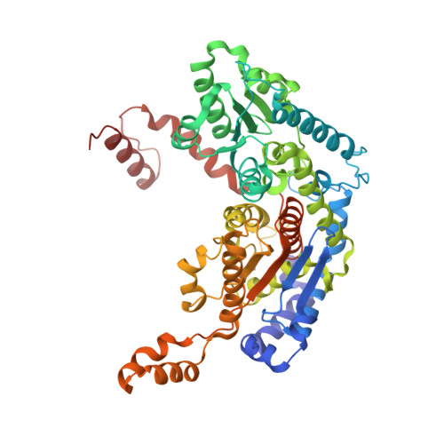
Conformational Changes in Phosphoglucose Isomerase Induced by Ligand Binding

Arsenieva, D.A.Jeffery, C.J.

(2002) J Mol Biology 323: 77-84

  • DOI: https://doi.org/10.1016/s0022-2836(02)00892-6
  • Primary Citation Related Structures: 
    1HM5

  • PubMed Abstract: 

    Phosphoglucose isomerase (PGI; EC 5.3.1.9) is the second enzyme in glycolysis, where it catalyzes the isomerization of D-glucose-6-phosphate to D-fructose-6-phosphate. It is the same protein as autocrine motility factor, differentiation and maturation mediator, and neuroleukin. Here, we report a new X-ray crystal structure of rabbit PGI (rPGI) without ligands bound in its active site. The structure was solved at 1.8A resolution by isomorphous phasing with a previously solved X-ray crystal structure of the rPGI dimer containing 6-phosphogluconate in its active site. Comparison of the new structure to previously reported structures enables identification of conformational changes that occur during binding of substrate or inhibitor molecules. Ligand binding causes an induced fit of regions containing amino acid residues 209-215, 245-259 and 385-389. This conformational change differs from the change previously reported to occur between the ring-opening and isomerization steps, in which the helix containing residues 513-521 moves toward the bound substrate. Differences between the liganded and unliganded structures are limited to the region within and close to the active-site pocket.


  • Organizational Affiliation
    • Laboratory for Molecular Biology, MC567, Department of Biological Sciences, University of Illinois, Chicago, IL 60607, USA.

Macromolecule Content 

  • Total Structure Weight: 125.66 kDa 
  • Atom Count: 9,740 
  • Modeled Residue Count: 1,112 
  • Deposited Residue Count: 1,116 
  • Unique protein chains: 1

Macromolecules

Find similar proteins by:|  3D Structure
Entity ID: 1
MoleculeChains  Sequence LengthOrganismDetailsImage
PHOSPHOGLUCOSE ISOMERASE
A, B
558Oryctolagus cuniculusMutation(s): 0 
EC: 5.3.1.9 (PDB Primary Data), 5.1.3 (UniProt)
UniProt
Find proteins for Q9N1E2 (Oryctolagus cuniculus)
Explore Q9N1E2 
Go to UniProtKB:  Q9N1E2
Entity Groups
Sequence Clusters30% Identity50% Identity70% Identity90% Identity95% Identity100% Identity
UniProt GroupQ9N1E2
Sequence Annotations
Expand
Reference Sequence

Experimental Data & Validation

Experimental Data

  • Method: X-RAY DIFFRACTION
  • Resolution: 1.80 Å
  • R-Value Free:  0.212 (Depositor), 0.212 (DCC) 
  • R-Value Work:  0.183 (Depositor), 0.183 (DCC) 
  • R-Value Observed: 0.183 (Depositor) 
Space Group: C 2 2 21
Unit Cell:
Length ( Å )Angle ( ˚ )
a = 82.756α = 90
b = 115.966β = 90
c = 271.845γ = 90
Software Package:
Software NamePurpose
DENZOdata reduction
SCALEPACKdata scaling
CNSrefinement
CNSphasing

Structure Validation

View Full Validation Report


Entry History 

Deposition Data

Revision History  (Full details and data files)

  • Version 1.0: 2002-09-27
    Type: Initial release
  • Version 1.1: 2008-04-27
    Changes: Version format compliance
  • Version 1.2: 2011-07-13
    Changes: Version format compliance
  • Version 1.3: 2011-11-16
    Changes: Atomic model
  • Version 1.4: 2023-08-09
    Changes: Data collection, Database references, Refinement description