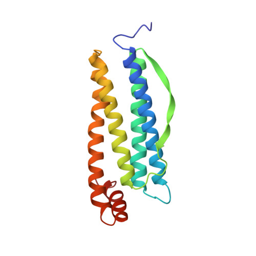
A crystallographic study of haem binding to ferritin.
Precigoux, G., Yariv, J., Gallois, B., Dautant, A., Courseille, C., d'Estaintot, B.L.(1994) Acta Crystallogr D Biol Crystallogr 50: 739-743
- PubMed: 15299370
- DOI: https://doi.org/10.1107/S0907444994003227
- Primary Citation of Related Structures:
1HRS - PubMed Abstract:
Ferritin, the iron-storage protein, binds porphyrins, metalloporphyrins and the fluorescent dyes ANS (8-anilino-1-naphthalenesulfonic acid) and TNS (2-p-toluidinyl-6-naphthalenesulfonic acid), similarly to apo-myoglobin. Octahedral crystals of horse-spleen apo-ferritin (HSF; 174 amino acids) complexes prepared by the addition of haem, hematoporphyrin or Sn-protoporphyrin IX to a solution of apo-ferritin crystallize in space group F432 with cell parameter a = 184.0 A. X-ray crystallographic analysis of single crystals prepared from a mixture containing haem or Sn-protoporphyrin IX shows that the haem-binding sites in these crystals are occupied by protoporphyrin IX, which is free of metal, rather than by the original metalloporphyrin. The present paper describes the structure of horse-spleen apo-ferritin cocrystallized with Sn-protoporphyrin IX. The 6797 reflections up to 2.6 A resolution used in the refinement were obtained from a data set recorded on a Nicolet/Xentronics area detector with Cu Kalpha radiation from a Rigaku RU 200 rotating anode. The final structure comprises 1613 non-H atoms, two Cd atoms and 170 solvent molecules. Four residues are described as disordered. The root-mean-square deviations from ideal bond lengths and angles are 0.013 A and 2.88 degrees, respectively. Protoporphyrins are observed in special positions on the twofold axes of the ferritin molecule with a stoichiometry of 0.4 per subunit.
- Laboratoire de Cristallographie et Physique Cristalline, Université de Bordeaux, Talence, France.
Organizational Affiliation:


