1I2H | pdb_00001i2h

CRYSTAL STRUCTURE ANALYSIS OF PSD-ZIP45(HOMER1C/VESL-1L)CONSERVED HOMER 1 DOMAIN


Experimental Data Snapshot

  • Method: X-RAY DIFFRACTION
  • Resolution: 1.80 Å
  • R-Value Free: 
    0.218 (Depositor), 0.212 (DCC) 
  • R-Value Work: 
    0.195 (Depositor), 0.207 (DCC) 
  • R-Value Observed: 
    0.223 (Depositor) 

Starting Model: experimental
View more details

wwPDB Validation 3D Report Full Report

Validation slider image for 1I2H

This is version 1.3 of the entry. See complete history

Literature

Crystal Structure of the Homer 1 Family Conserved Region Reveals the Interaction Between the EVH1 Domain and Own Proline-rich Motif

Irie, K.Nakatsu, T.Mitsuoka, K.Miyazawa, A.Sobue, K.Hiroaki, Y.Doi, T.Fujiyoshi, Y.Kato, H.

(2002) J Mol Biology 318: 1117-1126

  • DOI: https://doi.org/10.1016/S0022-2836(02)00170-5
  • Primary Citation Related Structures: 
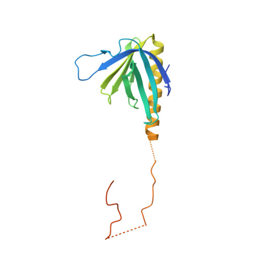
    1I2H

  • PubMed Abstract: 

    PSD-Zip45 (also named Homer 1c/Vesl-1L) is a synaptic scaffolding protein, which interacts with neurotransmitter receptors and other scaffolding proteins to target them into post-synaptic density (PSD), a specialized protein complex at the synaptic junction. Binding of the PSD-Zip45 to the receptors and scaffolding proteins results in colocalization and clustering of its binding partners in PSD. It has an Ena/VASP homology 1 (EVH1) domain in the N terminus for receptor binding, two leucine zipper motifs in the C terminus for clustering, and a linking region whose function is unclear despite the high level of conservation within the Homer 1 family. The X-ray crystallographic analysis of the largest fragment of residues 1-163, including an EVH1 domain reported here, demonstrates that the EVH1 domain contains an alpha-helix longer than that of the previous models, and that the linking part included in the conserved region of Homer 1 (CRH1) of the PSD-Zip45 interacts with the EVH1 domain of the neighbour CRH1 molecule in the crystal. The results suggest that the EVH1 domain recognizes the PPXXF motif found in the binding partners, and the SPLTP sequence (P-motif) in the linking region of the CRH1. The two types of binding are partly overlapped in the EVH1 domain, implying a mechanism to regulate multimerization of Homer 1 family proteins.


  • Organizational Affiliation
    • Department of Biophysics, Kyoto University Graduate School of Science, Oiwake-cho, Kitashirakawa, Sakyo-ku, 606-8502, Japan.

Macromolecule Content 

  • Total Structure Weight: 18.82 kDa 
  • Atom Count: 1,170 
  • Modeled Residue Count: 138 
  • Deposited Residue Count: 168 
  • Unique protein chains: 1

Macromolecules

Find similar proteins by:|  3D Structure
Entity ID: 1
MoleculeChains  Sequence LengthOrganismDetailsImage
PSD-ZIP45(HOMER-1C/VESL-1L)168Rattus norvegicusMutation(s): 0 
Gene Names: PSD-ZIP45
UniProt
Find proteins for Q9Z214 (Rattus norvegicus)
Explore Q9Z214 
Go to UniProtKB:  Q9Z214
Entity Groups
Sequence Clusters30% Identity50% Identity70% Identity90% Identity95% Identity100% Identity
UniProt GroupQ9Z214
Sequence Annotations
Expand
Reference Sequence

Experimental Data & Validation

Experimental Data

  • Method: X-RAY DIFFRACTION
  • Resolution: 1.80 Å
  • R-Value Free:  0.218 (Depositor), 0.212 (DCC) 
  • R-Value Work:  0.195 (Depositor), 0.207 (DCC) 
  • R-Value Observed: 0.223 (Depositor) 
Space Group: P 21 21 2
Unit Cell:
Length ( Å )Angle ( ˚ )
a = 55.262α = 90
b = 71.153β = 90
c = 39.565γ = 90
Software Package:
Software NamePurpose
AMoREphasing
REFMACrefinement
MOSFLMdata reduction
CCP4data scaling

Structure Validation

View Full Validation Report


Entry History 

Deposition Data

Revision History  (Full details and data files)

  • Version 1.0: 2002-05-29
    Type: Initial release
  • Version 1.1: 2008-04-27
    Changes: Version format compliance
  • Version 1.2: 2011-07-13
    Changes: Version format compliance
  • Version 1.3: 2023-08-09
    Changes: Data collection, Database references, Refinement description