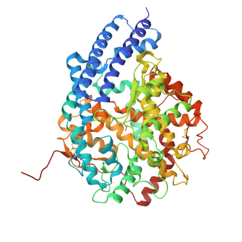
1J38

Crystal Structure of Drosophila AnCE


Experimental Data Snapshot

  • Method: X-RAY DIFFRACTION
  • Resolution: 2.60 Å
  • R-Value Free: 0.281 
  • R-Value Work: 0.246 
  • R-Value Observed: 0.246 

wwPDB Validation   3D Report Full Report


This is version 1.4 of the entry. See complete history


Literature

Crystal structure of Drosophila angiotensin I-converting enzyme bound to captopril and lisinopril

Kim, H.M.Shin, D.R.Yoo, O.J.Lee, H.Lee, J.-O.

(2003) FEBS Lett 538: 65-70

  • DOI: https://doi.org/10.1016/s0014-5793(03)00128-5
  • Primary Citation of Related Structures:  
    1J36, 1J37, 1J38

  • PubMed Abstract: 

    Angiotensin I-converting enzymes (ACEs) are zinc metallopeptidases that cleave carboxy-terminal dipeptides from short peptide hormones. We have determined the crystal structures of AnCE, a Drosophila homolog of ACE, with and without bound inhibitors to 2.4 A resolution. AnCE contains a large internal channel encompassing the entire protein molecule. This substrate-binding channel is composed of two chambers, reminiscent of a peanut shell. The inhibitor and zinc-binding sites are located in the narrow bottleneck connecting the two chambers. The substrate and inhibitor specificity of AnCE appears to be determined by extensive hydrogen-bonding networks and ionic interactions in the active site channel. The catalytically important zinc ion is coordinated by the conserved Glu395 and histidine residues from a HExxH motif.


  • Organizational Affiliation

    Department of Biological Science, Korea Advanced Institute of Science and Technology, 373-1 Kusong-dong, Yusong-gu, Daejeon, South Korea.


Macromolecules
Find similar proteins by:  (by identity cutoff)  |  3D Structure
Entity ID: 1
MoleculeChains Sequence LengthOrganismDetailsImage
angiotensin converting enzyme
A, B
607Drosophila melanogasterMutation(s): 0 
EC: 3.4.15.1
UniProt
Find proteins for Q10714 (Drosophila melanogaster)
Explore Q10714 
Go to UniProtKB:  Q10714
Entity Groups  
Sequence Clusters30% Identity50% Identity70% Identity90% Identity95% Identity100% Identity
UniProt GroupQ10714
Sequence Annotations
Expand
  • Reference Sequence
Experimental Data & Validation

Experimental Data

  • Method: X-RAY DIFFRACTION
  • Resolution: 2.60 Å
  • R-Value Free: 0.281 
  • R-Value Work: 0.246 
  • R-Value Observed: 0.246 
  • Space Group: P 1 21 1
Unit Cell:
Length ( Å )Angle ( ˚ )
a = 94.912α = 90
b = 121.223β = 99.39
c = 94.74γ = 90
Software Package:
Software NamePurpose
DENZOdata reduction
SCALEPACKdata scaling
SOLVEphasing
CNSrefinement

Structure Validation

View Full Validation Report


Entry History 

Deposition Data

Revision History  (Full details and data files)

  • Version 1.0: 2003-07-20
    Type: Initial release
  • Version 1.1: 2008-04-27
    Changes: Version format compliance
  • Version 1.2: 2011-07-13
    Changes: Version format compliance
  • Version 1.3: 2023-12-27
    Changes: Data collection, Database references, Derived calculations
  • Version 1.4: 2024-10-16
    Changes: Structure summary