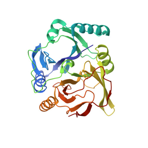
Crystal Structures of the Reaction Intermediate and its Homologue of an Extradiol-cleaving Catecholic Dioxygenase
Sato, N., Uragami, Y., Nishizaki, T., Takahashi, Y., Sazaki, G., Sugimoto, K., Nonaka, T., Masai, E., Fukuda, M., Senda, T.(2002) J Mol Biology 321: 621-636
- PubMed: 12206778
- DOI: https://doi.org/10.1016/s0022-2836(02)00673-3
- Primary Citation of Related Structures:
1KW3, 1KW6, 1KW8, 1KW9, 1KWB, 1KWC - PubMed Abstract:
BphC derived from Pseudomonas sp. strain KKS102 is an extradiol-cleaving catecholic dioxygenase. This enzyme contains a non-heme iron atom and plays an important role in degrading biphenyl/polychlorinated biphenyls (PCBs) in the microbe. To elucidate detailed structures of BphC reaction intermediates, crystal structures of the substrate-free form, the BphC-substrate complex, and the BphC-substrate-NO (nitric oxide) complex were determined. These crystal structures revealed (1) the binding site of the O(2) molecule in the coordination sphere and (2) conformational changes of His194 during the catalytic reaction. On the basis of these findings, we propose a catalytic mechanism for the extradiol-cleaving catecholic dioxygenase in which His194 seems to play three distinct roles. At the early stage of the catalytic reaction, His194 appears to act as a catalytic base, which likely deprotonates the hydroxyl group of the substrate. At the next stage, the protonated His194 seems to stabilize a negative charge on the O2 molecule located in the hydrophobic O2-binding cavity. Finally, protonated His194 seems to function as a proton donor, whose existence has been proposed.
- Department of BioEngineering, Nagaoka University of Technology, Nagaoka, Niigata, Japan.
Organizational Affiliation:


