1KY9 | pdb_00001ky9

Crystal Structure of DegP (HtrA)


Experimental Data Snapshot

  • Method: X-RAY DIFFRACTION
  • Resolution: 2.80 Å
  • R-Value Free: 
    0.275 (Depositor), 0.286 (DCC) 
  • R-Value Work: 
    0.218 (Depositor), 0.228 (DCC) 
  • R-Value Observed: 
    0.226 (Depositor) 

wwPDB Validation 3D Report Full Report

Validation slider image for 1KY9

This is version 1.4 of the entry. See complete history

Literature

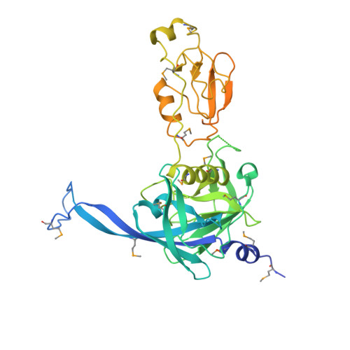
Crystal structure of DegP (HtrA) reveals a new protease-chaperone machine.

Krojer, T.Garrido-Franco, M.Huber, R.Ehrmann, M.Clausen, T.

(2002) Nature 416: 455-459

  • DOI: https://doi.org/10.1038/416455a
  • Primary Citation Related Structures: 
    1KY9

  • PubMed Abstract: 

    Molecular chaperones and proteases monitor the folded state of other proteins. In addition to recognizing non-native conformations, these quality control factors distinguish substrates that can be refolded from those that need to be degraded. To investigate the molecular basis of this process, we have solved the crystal structure of DegP (also known as HtrA), a widely conserved heat shock protein that combines refolding and proteolytic activities. The DegP hexamer is formed by staggered association of trimeric rings. The proteolytic sites are located in a central cavity that is only accessible laterally. The mobile side-walls are constructed by twelve PDZ domains, which mediate the opening and closing of the particle and probably the initial binding of substrate. The inner cavity is lined by several hydrophobic patches that may act as docking sites for unfolded polypeptides. In the chaperone conformation, the protease domain of DegP exists in an inactive state, in which substrate binding in addition to catalysis is abolished.


  • Organizational Affiliation
    • Max-Planck-Institut für Biochemie, Abteilung Strukturforschung, Am Klopferspitz 18A, 82152 Martinsried, Germany.

Macromolecule Content 

  • Total Structure Weight: 95.02 kDa 
  • Atom Count: 5,366 
  • Modeled Residue Count: 707 
  • Deposited Residue Count: 896 
  • Unique protein chains: 1

Macromolecules

Find similar proteins by:|  3D Structure
Entity ID: 1
MoleculeChains  Sequence LengthOrganismDetailsImage
PROTEASE DO
A, B
448Escherichia coliMutation(s): 15 
Gene Names: degP
EC: 3.4.21 (PDB Primary Data), 3.4.21.107 (UniProt)
UniProt
Find proteins for P0C0V0 (Escherichia coli (strain K12))
Explore P0C0V0 
Go to UniProtKB:  P0C0V0
Entity Groups
Sequence Clusters30% Identity50% Identity70% Identity90% Identity95% Identity100% Identity
UniProt GroupP0C0V0
Sequence Annotations
Expand
Reference Sequence

Small Molecules

Modified Residues  1 Unique
IDChains TypeFormula2D DiagramParent
MSE
Query on MSE
A, B
L-PEPTIDE LINKINGC5 H11 N O2 SeMET

Experimental Data & Validation

Experimental Data

  • Method: X-RAY DIFFRACTION
  • Resolution: 2.80 Å
  • R-Value Free:  0.275 (Depositor), 0.286 (DCC) 
  • R-Value Work:  0.218 (Depositor), 0.228 (DCC) 
  • R-Value Observed: 0.226 (Depositor) 
Space Group: P 63 2 2
Unit Cell:
Length ( Å )Angle ( ˚ )
a = 121.374α = 90
b = 121.374β = 90
c = 233.667γ = 120
Software Package:
Software NamePurpose
SHARPphasing
CNSrefinement
DENZOdata reduction
SCALEPACKdata scaling

Structure Validation

View Full Validation Report


Entry History 

Deposition Data

Revision History  (Full details and data files)

  • Version 1.0: 2002-04-03
    Type: Initial release
  • Version 1.1: 2008-04-28
    Changes: Version format compliance
  • Version 1.2: 2011-07-13
    Changes: Version format compliance
  • Version 1.3: 2021-10-27
    Changes: Database references, Derived calculations
  • Version 1.4: 2024-11-13
    Changes: Data collection, Structure summary