1KZC

Complex of MBP-C and high-affinity linear trimannose


Experimental Data Snapshot

  • Method: X-RAY DIFFRACTION
  • Resolution: 1.85 Å
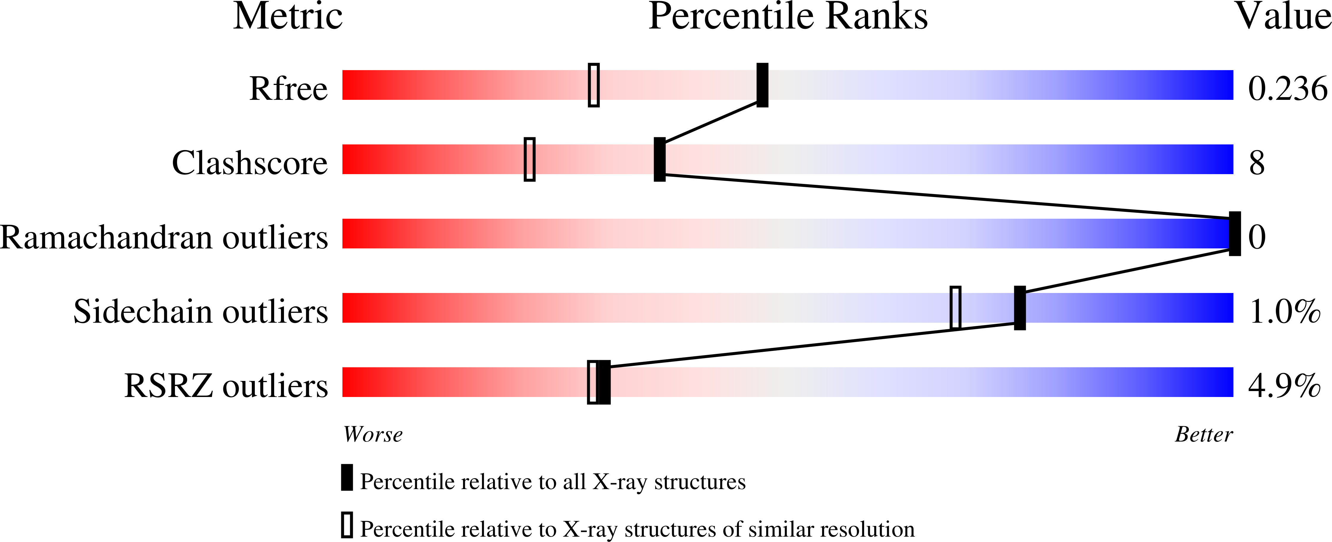
  • R-Value Free: 0.242 
  • R-Value Work: 0.210 
  • R-Value Observed: 0.213 

Starting Model: experimental
View more details

wwPDB Validation   3D Report Full Report


Ligand Structure Quality Assessment 


This is version 1.6 of the entry. See complete history


Literature

Orientation of bound ligands in mannose-binding proteins. Implications for multivalent ligand recognition.

Ng, K.K.Kolatkar, A.R.Park-Snyder, S.Feinberg, H.Clark, D.A.Drickamer, K.Weis, W.I.

(2002) J Biol Chem 277: 16088-16095

  • DOI: https://doi.org/10.1074/jbc.M200493200
  • Primary Citation of Related Structures:  
    1KWT, 1KWU, 1KWV, 1KWW, 1KWX, 1KWY, 1KWZ, 1KX0, 1KX1, 1KZA, 1KZB, 1KZC, 1KZD, 1KZE

  • PubMed Abstract: 

    Mannose-binding proteins (MBPs) are C-type animal lectins that recognize high mannose oligosaccharides on pathogenic cell surfaces. MBPs bind to their carbohydrate ligands by forming a series of Ca(2+) coordination and hydrogen bonds with two hydroxyl groups equivalent to the 3- and 4-OH of mannose. In this work, the determinants of the orientation of sugars bound to rat serum and liver MBPs (MBP-A and MBP-C) have been systematically investigated. The crystal structures of MBP-A soaked with monosaccharides and disaccharides and also the structure of the MBP-A trimer cross-linked by a high mannose asparaginyl oligosaccharide reveal that monosaccharides or alpha1-6-linked mannose bind to MBP-A in one orientation, whereas alpha1-2- or alpha1-3-linked mannose binds in an orientation rotated 180 degrees around a local symmetry axis relating the 3- and 4-OH groups. In contrast, a similar set of ligands all bind to MBP-C in a single orientation. The mutation of MBP-A His(189) to its MBP-C equivalent, valine, causes Man alpha 1-3Man to bind in a mixture of orientations. These data combined with modeling indicate that the residue at this position influences the orientation of bound ligands in MBP. We propose that the control of binding orientation can influence the recognition of multivalent ligands. A lateral association of trimers in the cross-linked crystals may reflect interactions within higher oligomers of MBP-A that are stabilized by multivalent ligands.


  • Organizational Affiliation

    Department of Structural Biology, Stanford University School of Medicine, Stanford, California 94305, USA.


Macromolecules
Find similar proteins by:  (by identity cutoff)  |  3D Structure
Entity ID: 1
MoleculeChains Sequence LengthOrganismDetailsImage
MANNOSE-BINDING PROTEIN CA [auth 1],
B [auth 2]
115Rattus norvegicusMutation(s): 0 
Gene Names: MBL1
UniProt
Find proteins for P08661 (Rattus norvegicus)
Explore P08661 
Go to UniProtKB:  P08661
Entity Groups  
Sequence Clusters30% Identity50% Identity70% Identity90% Identity95% Identity100% Identity
UniProt GroupP08661
Sequence Annotations
Expand
  • Reference Sequence
Small Molecules
Ligands 3 Unique
IDChains Name / Formula / InChI Key2D Diagram3D Interactions
MAN
Query on MAN

Download Ideal Coordinates CCD File 
C [auth 1],
G [auth 2]
alpha-D-mannopyranose
C6 H12 O6
WQZGKKKJIJFFOK-PQMKYFCFSA-N
CA
Query on CA

Download Ideal Coordinates CCD File 
D [auth 1],
E [auth 1],
H [auth 2],
I [auth 2]
CALCIUM ION
Ca
BHPQYMZQTOCNFJ-UHFFFAOYSA-N
CL
Query on CL

Download Ideal Coordinates CCD File 
F [auth 1]CHLORIDE ION
Cl
VEXZGXHMUGYJMC-UHFFFAOYSA-M
Experimental Data & Validation

Experimental Data

  • Method: X-RAY DIFFRACTION
  • Resolution: 1.85 Å
  • R-Value Free: 0.242 
  • R-Value Work: 0.210 
  • R-Value Observed: 0.213 
  • Space Group: P 21 21 21
Unit Cell:
Length ( Å )Angle ( ˚ )
a = 60.8α = 90
b = 75.21β = 90
c = 57.4γ = 90
Software Package:
Software NamePurpose
DENZOdata reduction
SCALEPACKdata scaling
X-PLORmodel building
CNSrefinement
X-PLORphasing

Structure Validation

View Full Validation Report


Ligand Structure Quality Assessment 


Entry History 

Deposition Data

Revision History  (Full details and data files)

  • Version 1.0: 2002-07-05
    Type: Initial release
  • Version 1.1: 2008-04-28
    Changes: Version format compliance
  • Version 1.2: 2011-07-13
    Changes: Version format compliance
  • Version 1.3: 2018-04-04
    Changes: Data collection
  • Version 1.4: 2020-07-29
    Type: Remediation
    Reason: Carbohydrate remediation
    Changes: Data collection, Derived calculations, Structure summary
  • Version 1.5: 2023-08-16
    Changes: Data collection, Database references, Refinement description, Structure summary
  • Version 1.6: 2024-10-30
    Changes: Structure summary