1L4X | pdb_00001l4x

Octameric de novo designed peptide


Experimental Data Snapshot

  • Method: X-RAY DIFFRACTION
  • Resolution: 2.00 Å
  • R-Value Free: 
    0.278 (Depositor), 0.278 (DCC) 
  • R-Value Work: 
    0.242 (Depositor), 0.235 (DCC) 

wwPDB Validation 3D Report Full Report

Validation slider image for 1L4X

This is version 3.1 of the entry. See complete history

Literature

Removing an interhelical salt bridge abolishes coiled-coil formation in a de novo designed peptide

Meier, M.Lustig, A.Aebi, U.Burkhard, P.

(2002) J Struct Biol 137: 65-72

  • DOI: https://doi.org/10.1006/jsbi.2002.4467
  • Primary Citation Related Structures: 
    1L4X

  • PubMed Abstract: 

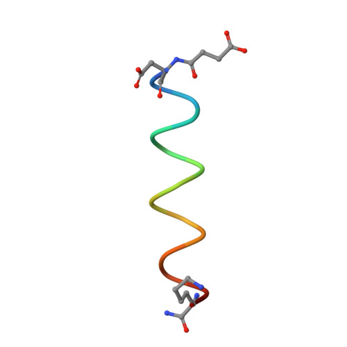
    Alpha-helical coiled coils represent a common protein oligomerization motif that are mainly stabilized by hydrophobic interactions occurring along their coiled-coil interface, the so-called hydrophobic seam. We have recently de novo designed and optimized a series of two-heptad repeat long coiled-coil peptides which are further stabilized by a complex network of inter- and intrahelical salt bridges. Here we have extended the de novo design of such two heptad-repeat long peptides by removing the central and most important g-e' Arg to Glu (g-e'RE) ionic interhelical interaction and replacing these residues by alanine residues. The effect of the missing interhelical ionic interaction on coiled-coil formation and stability has been analyzed by CD spectroscopy, analytical ultracentrifugation, and X-ray crystallography. We show that the peptide, while being highly alpha-helical, is no longer able to form a parallel coiled-coil structure but rather assumes an octameric globular helical assembly devoid of any coiled-coil interactions.


  • Organizational Affiliation
    • M.E. Müller Institute for Structural Biology, University of Basel, Switzerland.

Macromolecule Content 

  • Total Structure Weight: 15.14 kDa 
  • Atom Count: 1,155 
  • Modeled Residue Count: 128 
  • Deposited Residue Count: 128 
  • Unique protein chains: 1

Macromolecules

Find similar proteins by:  Sequence   |   3D Structure  
Entity ID: 1
MoleculeChains  Sequence LengthOrganismDetailsImage
SIN-ASP-GLU-LEU-GLU-ARG-ALA-ILE-ARG-GLU-LEU-ALA-ALA-ARG-ILE-LYS-NH2
A, B, C, D, E
A, B, C, D, E, F, G, H
16synthetic constructMutation(s): 0 

Small Molecules

Modified Residues  1 Unique
IDChains TypeFormula2D DiagramParent
ZJU
Query on ZJU
A, B, C, D, E
A, B, C, D, E, F, G, H
L-PEPTIDE LINKINGC8 H11 N O7ASP

Experimental Data & Validation

Experimental Data

  • Method: X-RAY DIFFRACTION
  • Resolution: 2.00 Å
  • R-Value Free:  0.278 (Depositor), 0.278 (DCC) 
  • R-Value Work:  0.242 (Depositor), 0.235 (DCC) 
Space Group: P 31 2 1
Unit Cell:
Length ( Å )Angle ( ˚ )
a = 56.628α = 90
b = 56.628β = 90
c = 113.869γ = 120
Software Package:
Software NamePurpose
DENZOdata reduction
TRUNCATEdata reduction
SHARPphasing
CNSrefinement
CCP4data scaling

Structure Validation

View Full Validation Report


Entry History 

Deposition Data

Revision History  (Full details and data files)

  • Version 1.0: 2002-11-13
    Type: Initial release
  • Version 1.1: 2008-04-28
    Changes: Version format compliance
  • Version 1.2: 2011-07-13
    Changes: Version format compliance
  • Version 1.3: 2018-01-31
    Changes: Experimental preparation
  • Version 2.0: 2023-06-07
    Changes: Advisory, Atomic model, Data collection, Database references, Derived calculations, Polymer sequence, Source and taxonomy, Structure summary
  • Version 3.0: 2024-04-24
    Changes: Advisory, Atomic model, Data collection, Database references, Derived calculations, Non-polymer description, Polymer sequence, Source and taxonomy, Structure summary
  • Version 3.1: 2024-11-13
    Changes: Structure summary