1L5P | pdb_00001l5p

Crystal Structure of Trichomonas vaginalis Ferredoxin


Experimental Data Snapshot

  • Method: X-RAY DIFFRACTION
  • Resolution: 2.20 Å
  • R-Value Free: 
    0.315 (Depositor) 
  • R-Value Work: 
    0.248 (Depositor) 

wwPDB Validation 3D Report Full Report

Validation slider image for 1L5P

This is version 1.3 of the entry. See complete history

Literature

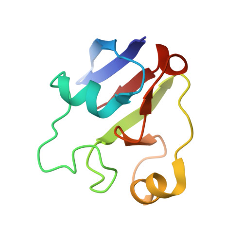
The crystal structure of Trichomonas vaginalis ferredoxin provides insight into metronidazole activation.

Crossnoe, C.R.Germanas, J.P.LeMagueres, P.Mustata, G.Krause, K.L.

(2002) J Mol Biology 318: 503-518

  • DOI: https://doi.org/10.1016/S0022-2836(02)00051-7
  • Primary Citation Related Structures: 
    1L5P

  • PubMed Abstract: 

    Crystallographic studies revealing the three-dimensional structure of the oxidized form of the [2Fe-2S] ferredoxin from Trichomonas vaginalis (TvFd) are presented. TvFd, a member of the hydrogenosomal class of ferredoxins, possesses a unique combination of redox and spectroscopic properties, and is believed to be the biological molecule that activates the drug metronidazole reductively in the treatment of trichomoniasis. It is the first hydrogenosomal ferredoxin to have its structure determined. The structure of TvFd reveals a monomeric, 93 residue protein with a fold similar to that of other known [2Fe-2S] ferredoxins. It contains nine hydrogen bonds to the sulfur atoms of the cluster, which is more than the number predicted on the basis of the spectroscopic data. The TvFd structure contains a large dipole moment like adrenodoxin, and appears to have a similar interaction domain. Our analysis demonstrates that TvFd has a unique cavity near the iron-sulfur cluster that exposes one of the inorganic sulfur atoms of the cluster to solvent. This cavity is not seen in any other [2Fe-2S] ferredoxin with known structure, and is hypothesized to be responsible for the high rate of metronidazole reduction by TvFd.


  • Organizational Affiliation
    • Structural and Computational Biology and Molecular Biophysics Program, Baylor College of Medicine, Houston, TX 77030, USA.

Macromolecule Content 

  • Total Structure Weight: 29.9 kDa 
  • Atom Count: 2,105 
  • Modeled Residue Count: 279 
  • Deposited Residue Count: 279 
  • Unique protein chains: 1

Macromolecules

Find similar proteins by:|  3D Structure
Entity ID: 1
MoleculeChains  Sequence LengthOrganismDetailsImage
ferredoxin
A, B, C
93Trichomonas vaginalisMutation(s): 0 
Gene Names: pET-3a-TvFd
UniProt
Find proteins for P21149 (Trichomonas vaginalis)
Explore P21149 
Go to UniProtKB:  P21149
Entity Groups
Sequence Clusters30% Identity50% Identity70% Identity90% Identity95% Identity100% Identity
UniProt GroupP21149
Sequence Annotations
Expand
Reference Sequence

Experimental Data & Validation

Experimental Data

  • Method: X-RAY DIFFRACTION
  • Resolution: 2.20 Å
  • R-Value Free:  0.315 (Depositor) 
  • R-Value Work:  0.248 (Depositor) 
Space Group: P 21 21 21
Unit Cell:
Length ( Å )Angle ( ˚ )
a = 35.4α = 90
b = 64.55β = 90
c = 133.88γ = 90
Software Package:
Software NamePurpose
SOLVEphasing
X-PLORrefinement
DENZOdata reduction
SCALEPACKdata scaling

Structure Validation

View Full Validation Report


Entry History 

Deposition Data

Revision History  (Full details and data files)

  • Version 1.0: 2002-08-21
    Type: Initial release
  • Version 1.1: 2008-04-28
    Changes: Version format compliance
  • Version 1.2: 2011-07-13
    Changes: Version format compliance
  • Version 1.3: 2024-02-14
    Changes: Data collection, Database references, Derived calculations