Stabilization of Escherichia coli ribonuclease HI by cavity-filling mutations within a hydrophobic core.
Ishikawa, K., Nakamura, H., Morikawa, K., Kanaya, S.(1993) Biochemistry 32: 6171-6178
- PubMed: 8390295 Search on PubMed
- Primary Citation Related Structures:
1LAV, 1LAW - PubMed Abstract:
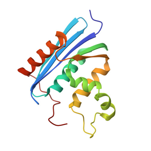
The crystal structure of Escherichia coli ribonuclease HI has a cavity near Val-74 within the protein core. In order to fill the cavity space, we constructed two mutant proteins, V74L and V74I, in which Val-74 was replaced with either Leu or Ile, respectively. The mutant proteins are stabilized, as revealed by a 2.1-3.7 degrees C increase in the Tm values, as compared to the wild-type protein at pH values of 3.0 and 5.5. The mutant protein V74A, in which Val-74 is replaced with Ala, was also constructed to analyze the reverse effect. The stability of V74A decreases by 7.6 degrees C at pH 3.0 and 12.7 degrees C at pH 5.5 in Tm as compared to those values for the wild-type protein. None of the three mutations significantly affect the enzymatic activity. The crystal structures of V74L and V74I, determined at 1.8-A resolution, are almost identical to that of the wild-type protein, except for the mutation site. In the two mutant proteins, calculation by the Voronoi procedure shows that the cavity volumes around the individual mutation sites are remarkably reduced as compared to that in the wild-type protein. These results indicate that the introduction of a methylene group into the cavity, without causing steric clash, contributes to an increase in the hydrophobic interaction within the protein core and thereby enhances protein stability. We also discuss the role of the Leu side chain, which can assume many different local conformations on a helix without sacrificing thermostability.
- Protein Engineering Research Institute, Osaka, Japan.
Organizational Affiliation: