1LRH

Crystal structure of auxin-binding protein 1 in complex with 1-naphthalene acetic acid


Experimental Data Snapshot

  • Method: X-RAY DIFFRACTION
  • Resolution: 1.90 Å
  • R-Value Free: 
    0.240 (Depositor), 0.240 (DCC) 
  • R-Value Work: 
    0.200 (Depositor), 0.190 (DCC) 
  • R-Value Observed: 
    0.200 (Depositor) 

wwPDB Validation   3D Report Full Report


Ligand Structure Quality Assessment 

Created with Raphaël 2.3.0Worse 01 BetterLigand structure goodness of fit to experimental dataBest fitted NLAClick on this verticalbar to view details

This is version 2.2 of the entry. See complete history


Literature

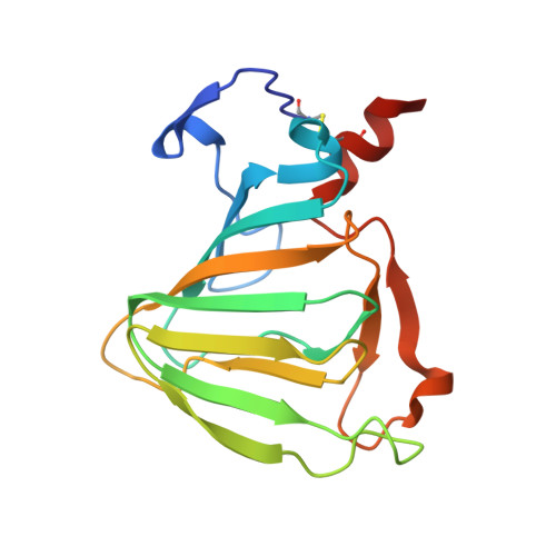
Crystal structure of auxin-binding protein 1 in complex with auxin.

Woo, E.J.Marshall, J.Bauly, J.Chen, J.G.Venis, M.Napier, R.M.Pickersgill, R.W.

(2002) EMBO J 21: 2877-2885

  • DOI: https://doi.org/10.1093/emboj/cdf291
  • Primary Citation of Related Structures:  
    1LR5, 1LRH

  • PubMed Abstract: 

    The structure of auxin-binding protein 1 (ABP1) from maize has been determined at 1.9 A resolution, revealing its auxin-binding site. The structure confirms that ABP1 belongs to the ancient and functionally diverse germin/seed storage 7S protein superfamily. The binding pocket of ABP1 is predominantly hydrophobic with a metal ion deep inside the pocket coordinated by three histidines and a glutamate. Auxin binds within this pocket, with its carboxylate binding the zinc and its aromatic ring binding hydrophobic residues including Trp151. There is a single disulfide between Cys2 and Cys155. No conformational rearrangement of ABP1 was observed when auxin bound to the protein in the crystal, but examination of the structure reveals a possible mechanism of signal transduction.


  • Organizational Affiliation

    Biological Sciences, Queen Mary, University of London, Mile End Road, London E1 4NS, UK.


Macromolecules
Find similar proteins by:  (by identity cutoff)  |  3D Structure
Entity ID: 1
MoleculeChains Sequence LengthOrganismDetailsImage
auxin-binding protein 1
A, B, C, D
163Zea maysMutation(s): 2 
Gene Names: ABP1
UniProt
Find proteins for P13689 (Zea mays)
Explore P13689 
Go to UniProtKB:  P13689
Entity Groups  
Sequence Clusters30% Identity50% Identity70% Identity90% Identity95% Identity100% Identity
UniProt GroupP13689
Glycosylation
Glycosylation Sites: 1
Sequence Annotations
Expand
  • Reference Sequence
Oligosaccharides

Help

Entity ID: 2
MoleculeChains Length2D Diagram Glycosylation3D Interactions
alpha-D-mannopyranose-(1-3)-[alpha-D-mannopyranose-(1-6)]alpha-D-mannopyranose-(1-6)-beta-D-mannopyranose-(1-4)-2-acetamido-2-deoxy-beta-D-glucopyranose-(1-4)-2-acetamido-2-deoxy-beta-D-glucopyranose
E, F, G, H
6N-Glycosylation
Glycosylation Resources
GlyTouCan:  G94106MV
GlyCosmos:  G94106MV
GlyGen:  G94106MV
Small Molecules
Ligands 2 Unique
IDChains Name / Formula / InChI Key2D Diagram3D Interactions
NLA
Query on NLA

Download Ideal Coordinates CCD File 
J [auth A],
L [auth B],
N [auth C],
P [auth D]
NAPHTHALEN-1-YL-ACETIC ACID
C12 H10 O2
PRPINYUDVPFIRX-UHFFFAOYSA-N
ZN
Query on ZN

Download Ideal Coordinates CCD File 
I [auth A],
K [auth B],
M [auth C],
O [auth D]
ZINC ION
Zn
PTFCDOFLOPIGGS-UHFFFAOYSA-N
Experimental Data & Validation

Experimental Data

  • Method: X-RAY DIFFRACTION
  • Resolution: 1.90 Å
  • R-Value Free:  0.240 (Depositor), 0.240 (DCC) 
  • R-Value Work:  0.200 (Depositor), 0.190 (DCC) 
  • R-Value Observed: 0.200 (Depositor) 
Space Group: P 1 21 1
Unit Cell:
Length ( Å )Angle ( ˚ )
a = 61.583α = 90
b = 82.433β = 94.37
c = 69.962γ = 90
Software Package:
Software NamePurpose
DENZOdata reduction
SCALEPACKdata scaling
REFMACrefinement

Structure Validation

View Full Validation Report


Ligand Structure Quality Assessment 

Created with Raphaël 2.3.0Worse 01 BetterLigand structure goodness of fit to experimental dataBest fitted NLAClick on this verticalbar to view details

Entry History 

Deposition Data

Revision History  (Full details and data files)

  • Version 1.0: 2002-06-19
    Type: Initial release
  • Version 1.1: 2008-04-28
    Changes: Version format compliance
  • Version 1.2: 2011-07-13
    Changes: Non-polymer description, Version format compliance
  • Version 1.3: 2017-10-11
    Changes: Refinement description
  • Version 2.0: 2020-07-29
    Type: Remediation
    Reason: Carbohydrate remediation
    Changes: Advisory, Atomic model, Data collection, Derived calculations, Structure summary
  • Version 2.1: 2021-11-10
    Changes: Database references, Structure summary
  • Version 2.2: 2024-11-13
    Changes: Data collection, Structure summary