1MNM | pdb_00001mnm

YEAST MATALPHA2/MCM1/DNA TERNARY TRANSCRIPTION COMPLEX CRYSTAL STRUCTURE


Experimental Data Snapshot

  • Method: X-RAY DIFFRACTION
  • Resolution: 2.25 Å
  • R-Value Free: 
    0.285 (Depositor) 
  • R-Value Work: 
    0.240 (Depositor) 
  • R-Value Observed: 
    0.240 (Depositor) 

Starting Models: experimental
View more details

wwPDB Validation 3D Report Full Report

Validation slider image for 1MNM

This is version 1.4 of the entry. See complete history

Literature

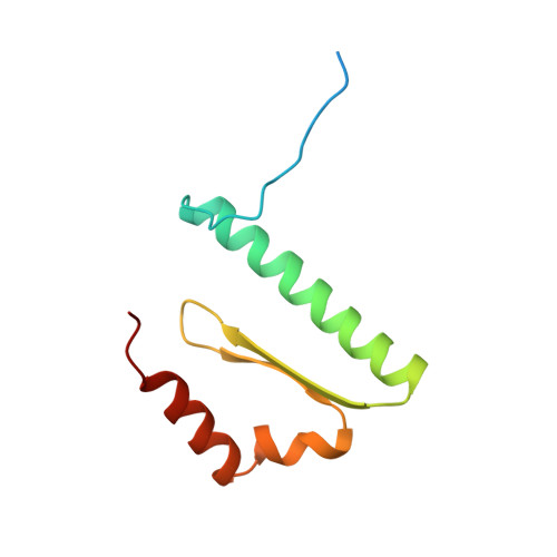
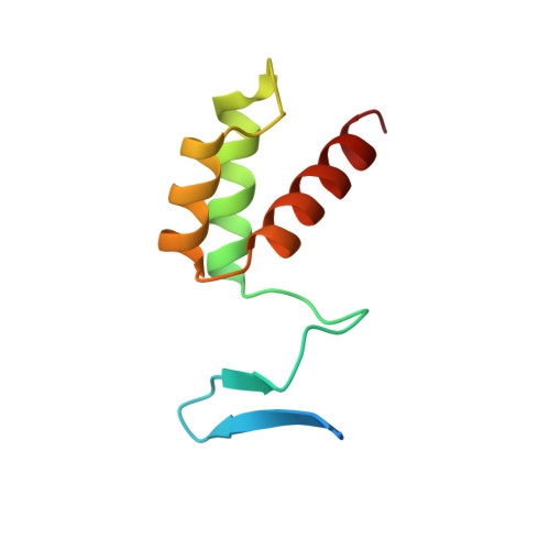
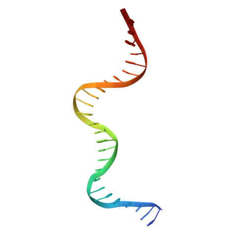
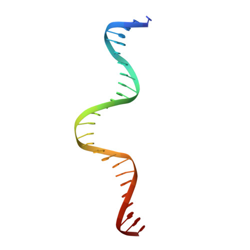
Crystal structure of the yeast MATalpha2/MCM1/DNA ternary complex.

Tan, S.Richmond, T.J.

(1998) Nature 391: 660-666

  • DOI: https://doi.org/10.1038/35563
  • Primary Citation Related Structures: 
    1MNM

  • PubMed Abstract: 

    The structure of a complex containing the homeodomain repressor protein MATalpha2 and the MADS-box transcription factor MCM1 bound to DNA has been determined by X-ray crystallography at 2.25 A resolution. It reveals the protein-protein interactions responsible for cooperative binding of MATalpha2 and MCM1 to DNA. The otherwise flexible amino-terminal extension of the MATalpha2 homeodomain forms a beta-hairpin that grips the MCM1 surface through parallel beta-strand hydrogen bonds and close-packed, predominantly hydrophobic, side chains. DNA bending induced by MCM1 brings the two proteins closer together, facilitating their interaction. An unusual feature of the complex is that an eight-amino-acid sequence adopts an alpha-helical conformation in one of two copies of the MATalpha2 monomer and a beta-strand conformation in the other. This 'chameleon' sequence of MATalpha2 may be important for recognizing natural operator sites.


  • Organizational Affiliation
    • ETH-Zurich, Institut für Molekularbiologie und Biophysik, Switzerland.

Macromolecule Content 

  • Total Structure Weight: 59.45 kDa 
  • Atom Count: 3,657 
  • Modeled Residue Count: 372 
  • Deposited Residue Count: 426 
  • Unique protein chains: 2
  • Unique nucleic acid chains: 2

Macromolecules


Find similar proteins by:|  3D Structure
Entity ID: 3
MoleculeChains  Sequence LengthOrganismDetailsImage
PROTEIN (MCM1 TRANSCRIPTIONAL REGULATOR)C [auth A],
D [auth B]
100Saccharomyces cerevisiaeMutation(s): 0 
UniProt
Find proteins for P11746 (Saccharomyces cerevisiae (strain ATCC 204508 / S288c))
Explore P11746 
Go to UniProtKB:  P11746
Entity Groups
Sequence Clusters30% Identity50% Identity70% Identity90% Identity95% Identity100% Identity
UniProt GroupP11746
Sequence Annotations
Expand
Reference Sequence
Find similar proteins by:|  3D Structure
Entity ID: 4
MoleculeChains  Sequence LengthOrganismDetailsImage
PROTEIN (MAT ALPHA-2 TRANSCRIPTIONAL REPRESSOR)E [auth C],
F [auth D]
87Saccharomyces cerevisiaeMutation(s): 0 
UniProt
Find proteins for P0CY08 (Saccharomyces cerevisiae (strain ATCC 204508 / S288c))
Explore P0CY08 
Go to UniProtKB:  P0CY08
Entity Groups
Sequence Clusters30% Identity50% Identity70% Identity90% Identity95% Identity100% Identity
UniProt GroupP0CY08
Sequence Annotations
Expand
Reference Sequence
Find similar nucleic acids by:  (by identity cutoff) 
Entity ID: 1
MoleculeChains LengthOrganismImage
DNA (STE6 OPERATOR DNA)A [auth E]26synthetic construct
Sequence Annotations
Expand
Reference Sequence
Find similar nucleic acids by:  (by identity cutoff) 
Entity ID: 2
MoleculeChains LengthOrganismImage
DNA (STE6 OPERATOR DNA)B [auth F]26synthetic construct
Sequence Annotations
Expand
Reference Sequence

Experimental Data & Validation

Experimental Data

  • Method: X-RAY DIFFRACTION
  • Resolution: 2.25 Å
  • R-Value Free:  0.285 (Depositor) 
  • R-Value Work:  0.240 (Depositor) 
  • R-Value Observed: 0.240 (Depositor) 
Space Group: P 21 21 21
Unit Cell:
Length ( Å )Angle ( ˚ )
a = 70.62α = 90
b = 72.55β = 90
c = 150.7γ = 90
Software Package:
Software NamePurpose
AMoREphasing
X-PLORrefinement

Structure Validation

View Full Validation Report


Entry History 

Deposition Data

Revision History  (Full details and data files)

  • Version 1.0: 1998-03-18
    Type: Initial release
  • Version 1.1: 2008-05-22
    Changes: Version format compliance
  • Version 1.2: 2011-07-13
    Changes: Version format compliance
  • Version 1.3: 2017-02-15
    Changes: Source and taxonomy, Structure summary
  • Version 1.4: 2023-08-02
    Changes: Database references, Refinement description