1MP1 | pdb_00001mp1

Solution structure of the PWI motif from SRm160


Experimental Data Snapshot

  • Method: SOLUTION NMR
  • Conformers Calculated: 200 
  • Conformers Submitted: 20 
  • Selection Criteria: structures with the lowest energy 

wwPDB Validation 3D Report Full Report

Validation slider image for 1MP1

This is version 1.4 of the entry. See complete history

Literature

Structure and function of the PWI motif: a novel nucleic acid-binding domain that facilitates pre-mRNA processing.

Szymczyna, B.R.Bowman, J.McCracken, S.Pineda-Lucena, A.Lu, Y.Cox, B.Lambermon, M.Graveley, B.R.Arrowsmith, C.H.Blencowe, B.J.

(2003) Genes Dev 17: 461-475

  • DOI: https://doi.org/10.1101/gad.1060403
  • Primary Citation Related Structures: 
    1MP1

  • PubMed Abstract: 

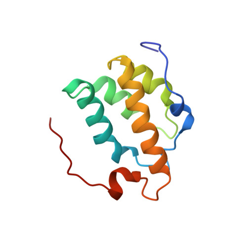
    The PWI motif is a highly conserved domain of unknown function in the SRm160 splicing and 3'-end cleavage-stimulatory factor, as well as in several other known or putative pre-mRNA processing components. We show here that the PWI motif is a new type of RNA/DNA-binding domain that has an equal preference for single- and double-stranded nucleic acids. Deletion of the motif prevents SRm160 from binding RNA and stimulating 3'-end cleavage, and its substitution with a heterologous RNA-binding domain restores these functions. The NMR solution structure of the SRm160-PWI motif reveals a novel, four-helix bundle and represents the first example of an alpha-helical fold that can bind single-stranded (ss)RNA. Structure-guided mutagenesis indicates that the same surface is involved in RNA and DNA binding and requires the cooperative action of a highly conserved, adjacent basic region. Thus, the PWI motif is a novel type of nucleic acid-binding domain that likely has multiple important functions in pre-mRNA processing, including SRm160-dependent stimulation of 3'-end formation.


  • Organizational Affiliation
    • Ontario Cancer Institute, University of Toronto, Toronto, Ontario, Canada M5G 2M9.

Macromolecule Content 

  • Total Structure Weight: 12.87 kDa 
  • Atom Count: 901 
  • Modeled Residue Count: 111 
  • Deposited Residue Count: 111 
  • Unique protein chains: 1

Macromolecules

Find similar proteins by:|  3D Structure
Entity ID: 1
MoleculeChains  Sequence LengthOrganismDetailsImage
Ser/Arg-related nuclear matrix protein111Homo sapiensMutation(s): 0 
Gene Names: SRm160
UniProt & NIH Common Fund Data Resources
Find proteins for Q8IYB3 (Homo sapiens)
Explore Q8IYB3 
Go to UniProtKB:  Q8IYB3
PHAROS:  Q8IYB3
GTEx:  ENSG00000133226 
Entity Groups
Sequence Clusters30% Identity50% Identity70% Identity90% Identity95% Identity100% Identity
UniProt GroupQ8IYB3
Sequence Annotations
Expand
Reference Sequence

Experimental Data & Validation

Experimental Data

  • Method: SOLUTION NMR
  • Conformers Calculated: 200 
  • Conformers Submitted: 20 
  • Selection Criteria: structures with the lowest energy 

Structure Validation

View Full Validation Report


Entry History 

Deposition Data

Revision History  (Full details and data files)

  • Version 1.0: 2003-09-16
    Type: Initial release
  • Version 1.1: 2008-04-28
    Changes: Version format compliance
  • Version 1.2: 2011-07-13
    Changes: Version format compliance
  • Version 1.3: 2022-02-23
    Changes: Database references, Derived calculations
  • Version 1.4: 2024-05-22
    Changes: Data collection