Download Mendeley
Crystallographic Characterization of an Exocyclic DNA Adduct: 3,N4-etheno-2'-deoxycytidine in the Dodecamer 5'-CGCGAATT(ethenoC)GCG-3'
Freisinger, E., Fernandes, A., Grollman, A.P., Kisker, C.F.(2003) J Mol Biol 329: 685-697
- PubMed: 12787670 Search on PubMed
- DOI: https://doi.org/10.1016/s0022-2836(03)00445-5
- Primary Citation Related Structures:
1N5C - PubMed Abstract:
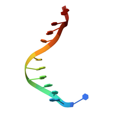
Exocyclic DNA adducts are formed from metabolites of chemical carcinogens and have also been detected as endogenous lesions in human DNA. The exocyclic adduct 3,N(4)-etheno-2'-deoxycytidine (epsilon dC), positioned opposite deoxyguanosine in the B-form duplex of the dodecanucleotide d(CGCGAATTepsilonCGCG), has been crystallographically characterized at 1.8A resolution. This self-complementary oligomer crystallizes in space group P3(2)12, containing a single strand in the asymmetric unit. The crystal structure was solved by isomorphous replacement with the corresponding unmodified dodecamer structure. Exposure of both structures to identical crystal packing forces allows a detailed investigation of the influence of the exocyclic base adduct on the overall helical structure and local geometry. Structural changes are limited to the epsilon C:G and adjacent T:A and G:C base-pairs. The standard Watson-Crick base-pairing scheme, retained in the T:A and G:C base-pairs, is blocked by the etheno bridge in the epsilon C:G pair. In its place, a hydrogen bond involving O2 of epsilon C and N1 of G is present. Comparison with an epsilon dC-containing NMR structure confirms the general conformation reported for epsilon C:G, including the hydrogen bonding features. Superposition with the crystal structure of a DNA duplex containing a T:G wobble pair shows similar structural changes imposed by both mismatches. Evaluation of the hydration shell of the duplex with bond valence calculations reveals two sodium ions in the crystal.
- Department of Pharmacological Sciences, Center for Structural Biology, State University of New York at Stony Brook, Stony Brook, NY 11794-5115, USA.
Organizational Affiliation: