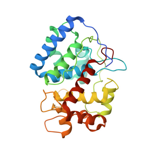
1OAF

Ascobate peroxidase from soybean cytosol in complex with ascorbate


Experimental Data Snapshot

  • Method: X-RAY DIFFRACTION
  • Resolution: 1.40 Å
  • R-Value Free: 0.196 
  • R-Value Work: 0.161 
  • R-Value Observed: 0.163 

Starting Model: experimental
View more details

wwPDB Validation   3D Report Full Report


This is version 1.4 of the entry. See complete history


Literature

Crystal Structure of the Ascorbate Peroxidase-Ascorbate Complex

Sharp, K.H.Mewies, M.Moody, P.C.E.Raven, E.L.

(2003) Nat Struct Biol 10: 303

  • DOI: https://doi.org/10.1038/nsb913
  • Primary Citation of Related Structures:  
    1OAF, 1OAG

  • PubMed Abstract: 

    Heme peroxidases catalyze the H2O2-dependent oxidation of a variety of substrates, most of which are organic. Mechanistically, these enzymes are well characterized: they share a common catalytic cycle that involves formation of a two-electron, oxidized Compound I intermediate followed by two single-electron reduction steps by substrate. The substrate specificity is more diverse--most peroxidases oxidize small organic substrates, but there are prominent exceptions--and there is a notable absence of structural information for a representative peroxidase-substrate complex. Thus, the features that control substrate specificity remain undefined. We present the structure of the complex of ascorbate peroxidase-ascorbate. The structure defines the ascorbate-binding interaction for the first time and provides new rationalization of the unusual functional features of the related cytochrome c peroxidase enzyme, which has been a benchmark for peroxidase catalysis for more than 20 years. A new mechanism for electron transfer is proposed that challenges existing views of substrate oxidation in other peroxidases.


  • Organizational Affiliation

    Department of Chemistry, University of Leicester, University Road, Leicester, LE1 7RH, England, UK.


Macromolecules
Find similar proteins by:  (by identity cutoff)  |  3D Structure
Entity ID: 1
MoleculeChains Sequence LengthOrganismDetailsImage
ASCORBATE PEROXIDASE261Glycine maxMutation(s): 0 
EC: 1.11.1.11
UniProt
Find proteins for Q43758 (Glycine max)
Go to UniProtKB:  Q43758
Sequence Annotations
Expand
  • Reference Sequence
Small Molecules
Ligands 3 Unique
IDChains Name / Formula / InChI Key2D Diagram3D Interactions
HEM
Query on HEM

Download Ideal Coordinates CCD File 
B [auth A]PROTOPORPHYRIN IX CONTAINING FE
C34 H32 Fe N4 O4
KABFMIBPWCXCRK-RGGAHWMASA-L
ASC
Query on ASC

Download Ideal Coordinates CCD File 
D [auth A]ASCORBIC ACID
C6 H8 O6
CIWBSHSKHKDKBQ-JLAZNSOCSA-N
NA
Query on NA

Download Ideal Coordinates CCD File 
C [auth A]SODIUM ION
Na
FKNQFGJONOIPTF-UHFFFAOYSA-N
Experimental Data & Validation

Experimental Data

  • Method: X-RAY DIFFRACTION
  • Resolution: 1.40 Å
  • R-Value Free: 0.196 
  • R-Value Work: 0.161 
  • R-Value Observed: 0.163 
  • Space Group: P 42 21 2
Unit Cell:
Length ( Å )Angle ( ˚ )
a = 81.328α = 90
b = 81.328β = 90
c = 74.884γ = 90
Software Package:
Software NamePurpose
REFMACrefinement
DENZOdata reduction
SCALEPACKdata scaling
MOLREPphasing

Structure Validation

View Full Validation Report


Entry History 

Deposition Data

Revision History  (Full details and data files)

  • Version 1.0: 2003-03-20
    Type: Initial release
  • Version 1.1: 2011-05-08
    Changes: Version format compliance
  • Version 1.2: 2011-07-13
    Changes: Version format compliance
  • Version 1.3: 2020-07-29
    Type: Remediation
    Reason: Carbohydrate remediation
    Changes: Data collection, Derived calculations, Other
  • Version 1.4: 2023-12-13
    Changes: Data collection, Database references, Refinement description, Structure summary