1OGT

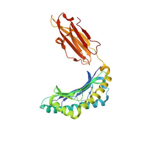
CRYSTAL STRUCTURE OF HLA-B*2705 COMPLEXED WITH THE VASOACTIVE INTESTINAL PEPTIDE TYPE 1 RECEPTOR (VIPR) PEPTIDE (RESIDUES 400-408)


Experimental Data Snapshot

  • Method: X-RAY DIFFRACTION
  • Resolution: 1.47 Å
  • R-Value Free: 0.178 
  • R-Value Work: 0.128 
  • R-Value Observed: 0.131 

Starting Model: experimental
View more details

wwPDB Validation   3D Report Full Report


This is version 2.3 of the entry. See complete history


Literature

Dual, HLA-B27 subtype-dependent conformation of a self-peptide.

Hulsmeyer, M.Fiorillo, M.T.Bettosini, F.Sorrentino, R.Saenger, W.Ziegler, A.Uchanska-Ziegler, B.

(2004) J Exp Med 199: 271-281

  • DOI: https://doi.org/10.1084/jem.20031690
  • Primary Citation of Related Structures:  
    1OF2, 1OGT

  • PubMed Abstract: 

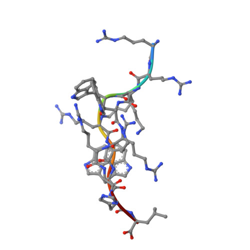
    The products of the human leukocyte antigen subtypes HLA-B*2705 and HLA-B*2709 differ only in residue 116 (Asp vs. His) within the peptide binding groove but are differentially associated with the autoimmune disease ankylosing spondylitis (AS); HLA-B*2705 occurs in AS-patients, whereas HLA-B*2709 does not. The subtypes also generate differential T cell repertoires as exemplified by distinct T cell responses against the self-peptide pVIPR (RRKWRRWHL). The crystal structures described here show that pVIPR binds in an unprecedented dual conformation only to HLA-B*2705 molecules. In one binding mode, peptide pArg5 forms a salt bridge to Asp116, connected with drastically different interactions between peptide and heavy chain, contrasting with the second, conventional conformation, which is exclusively found in the case of B*2709. These subtype-dependent differences in pVIPR binding link the emergence of dissimilar T cell repertoires in individuals with HLA-B*2705 or HLA-B*2709 to the buried Asp116/His116 polymorphism and provide novel insights into peptide presentation by major histocompatibility antigens.


  • Organizational Affiliation

    Institut für Kristallographie, Freie Universität Berlin, 14195 Berlin, Germany.


Macromolecules
Find similar proteins by:  (by identity cutoff)  |  3D Structure
Entity ID: 1
MoleculeChains Sequence LengthOrganismDetailsImage
HLA CLASS I HISTOCOMPATIBILITY ANTIGEN276Homo sapiensMutation(s): 0 
UniProt & NIH Common Fund Data Resources
Find proteins for P01889 (Homo sapiens)
Explore P01889 
Go to UniProtKB:  P01889
PHAROS:  P01889
GTEx:  ENSG00000234745 
Entity Groups  
Sequence Clusters30% Identity50% Identity70% Identity90% Identity95% Identity100% Identity
UniProt GroupP01889
Sequence Annotations
Expand
  • Reference Sequence
Find similar proteins by:  (by identity cutoff)  |  3D Structure
Entity ID: 2
MoleculeChains Sequence LengthOrganismDetailsImage
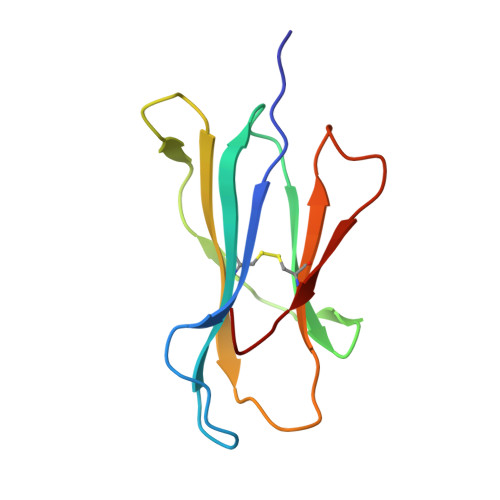
BETA-2-MICROGLOBULIN100Homo sapiensMutation(s): 0 
UniProt & NIH Common Fund Data Resources
Find proteins for P61769 (Homo sapiens)
Explore P61769 
Go to UniProtKB:  P61769
PHAROS:  P61769
GTEx:  ENSG00000166710 
Entity Groups  
Sequence Clusters30% Identity50% Identity70% Identity90% Identity95% Identity100% Identity
UniProt GroupP61769
Sequence Annotations
Expand
  • Reference Sequence

Find similar proteins by:  Sequence   |   3D Structure  

Entity ID: 3
MoleculeChains Sequence LengthOrganismDetailsImage
VASOACTIVE INTESTINAL POLYPEPTIDE RECEPTOR 19Homo sapiensMutation(s): 0 
UniProt & NIH Common Fund Data Resources
Find proteins for P32241 (Homo sapiens)
Explore P32241 
Go to UniProtKB:  P32241
PHAROS:  P32241
GTEx:  ENSG00000114812 
Entity Groups  
UniProt GroupP32241
Sequence Annotations
Expand
  • Reference Sequence
Experimental Data & Validation

Experimental Data

  • Method: X-RAY DIFFRACTION
  • Resolution: 1.47 Å
  • R-Value Free: 0.178 
  • R-Value Work: 0.128 
  • R-Value Observed: 0.131 
  • Space Group: P 1 21 1
Unit Cell:
Length ( Å )Angle ( ˚ )
a = 51.259α = 90
b = 81.777β = 107.61
c = 65.617γ = 90
Software Package:
Software NamePurpose
REFMACrefinement
DENZOdata reduction
SCALEPACKdata scaling
MOLREPphasing

Structure Validation

View Full Validation Report


Entry History 

Deposition Data

Revision History  (Full details and data files)

  • Version 1.0: 2004-01-29
    Type: Initial release
  • Version 1.1: 2011-05-08
    Changes: Version format compliance
  • Version 1.2: 2011-07-13
    Changes: Version format compliance
  • Version 2.0: 2018-12-19
    Changes: Atomic model, Data collection, Database references, Derived calculations
  • Version 2.1: 2019-05-08
    Changes: Data collection, Experimental preparation
  • Version 2.2: 2019-05-22
    Changes: Data collection, Experimental preparation
  • Version 2.3: 2023-12-13
    Changes: Data collection, Database references, Derived calculations, Other, Refinement description