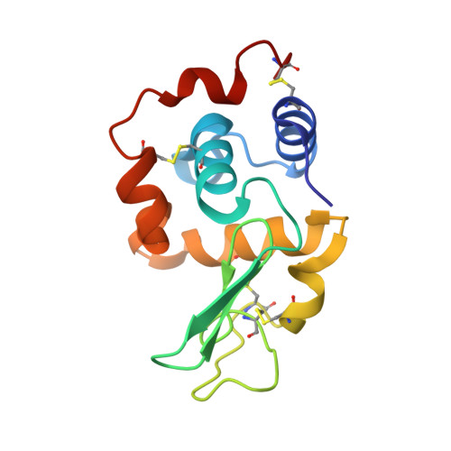
1OP9

Complex of human lysozyme with camelid VHH HL6 antibody fragment


Experimental Data Snapshot

  • Method: X-RAY DIFFRACTION
  • Resolution: 1.86 Å
  • R-Value Free: 0.238 
  • R-Value Work: 0.196 
  • R-Value Observed: 0.197 

Starting Models: experimental
View more details

wwPDB Validation   3D Report Full Report


This is version 1.5 of the entry. See complete history


Literature

A camelid antibody fragment inhibits the formation of amyloid fibrils by human lysozyme

Dumoulin, M.Last, A.M.Desmyter, A.Decanniere, K.Canet, D.Larsson, G.Spencer, A.Archer, D.B.Sasse, J.Muyldermans, S.Wyns, L.Redfield, C.Matagne, A.Robinson, C.V.Dobson, C.M.

(2003) Nature 424: 783-788

  • DOI: https://doi.org/10.1038/nature01870
  • Primary Citation of Related Structures:  
    1OP9

  • PubMed Abstract: 

    Amyloid diseases are characterized by an aberrant assembly of a specific protein or protein fragment into fibrils and plaques that are deposited in various organs and tissues, often with serious pathological consequences. Non-neuropathic systemic amyloidosis is associated with single point mutations in the gene coding for human lysozyme. Here we report that a single-domain fragment of a camelid antibody raised against wild-type human lysozyme inhibits the in vitro aggregation of its amyloidogenic variant, D67H. Our structural studies reveal that the epitope includes neither the site of mutation nor most residues in the region of the protein structure that is destabilized by the mutation. Instead, the binding of the antibody fragment achieves its effect by restoring the structural cooperativity characteristic of the wild-type protein. This appears to occur at least in part through the transmission of long-range conformational effects to the interface between the two structural domains of the protein. Thus, reducing the ability of an amyloidogenic protein to form partly unfolded species can be an effective method of preventing its aggregation, suggesting approaches to the rational design of therapeutic agents directed against protein deposition diseases.


  • Organizational Affiliation

    Department of Chemistry, University of Cambridge, Lensfield Road, Cambridge CB2 1EW, UK.


Macromolecules
Find similar proteins by:  (by identity cutoff)  |  3D Structure
Entity ID: 1
MoleculeChains Sequence LengthOrganismDetailsImage
HL6 camel VHH fragment121Camelus dromedariusMutation(s): 0 
Entity Groups  
Sequence Clusters30% Identity50% Identity70% Identity90% Identity95% Identity100% Identity
Sequence Annotations
Expand
  • Reference Sequence
Find similar proteins by:  (by identity cutoff)  |  3D Structure
Entity ID: 2
MoleculeChains Sequence LengthOrganismDetailsImage
Lysozyme C130Homo sapiensMutation(s): 0 
Gene Names: LYZ OR LZM
EC: 3.2.1.17
UniProt & NIH Common Fund Data Resources
Find proteins for P61626 (Homo sapiens)
Explore P61626 
Go to UniProtKB:  P61626
PHAROS:  P61626
GTEx:  ENSG00000090382 
Entity Groups  
Sequence Clusters30% Identity50% Identity70% Identity90% Identity95% Identity100% Identity
UniProt GroupP61626
Sequence Annotations
Expand
  • Reference Sequence
Experimental Data & Validation

Experimental Data

  • Method: X-RAY DIFFRACTION
  • Resolution: 1.86 Å
  • R-Value Free: 0.238 
  • R-Value Work: 0.196 
  • R-Value Observed: 0.197 
  • Space Group: P 1 21 1
Unit Cell:
Length ( Å )Angle ( ˚ )
a = 39.611α = 90
b = 48.226β = 98.98
c = 66.605γ = 90
Software Package:
Software NamePurpose
DENZOdata reduction
SCALAdata scaling
AMoREphasing
REFMACrefinement
CCP4data scaling

Structure Validation

View Full Validation Report


Entry History 

Revision History  (Full details and data files)

  • Version 1.0: 2003-10-14
    Type: Initial release
  • Version 1.1: 2008-04-29
    Changes: Version format compliance
  • Version 1.2: 2011-07-13
    Changes: Version format compliance
  • Version 1.3: 2018-04-04
    Changes: Data collection
  • Version 1.4: 2023-08-16
    Changes: Data collection, Database references, Refinement description
  • Version 1.5: 2024-11-06
    Changes: Structure summary