1OT5

The 2.4 Angstrom Crystal Structure of Kex2 in complex with a peptidyl-boronic acid inhibitor


Experimental Data Snapshot

  • Method: X-RAY DIFFRACTION
  • Resolution: 2.40 Å
  • R-Value Free: 
    0.245 (Depositor), 0.239 (DCC) 
  • R-Value Work: 
    0.209 (Depositor), 0.205 (DCC) 
  • R-Value Observed: 
    0.218 (Depositor) 

wwPDB Validation   3D Report Full Report


Ligand Structure Quality Assessment 

Created with Raphaël 2.3.0Worse 01 BetterLigand structure goodness of fit to experimental dataBest fitted NAGClick on this verticalbar to view details

This is version 2.1 of the entry. See complete history


Literature

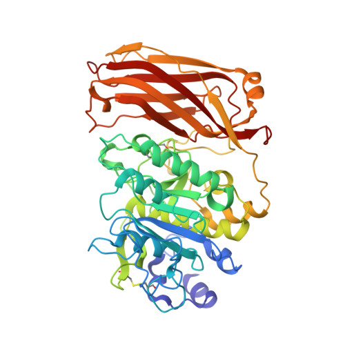
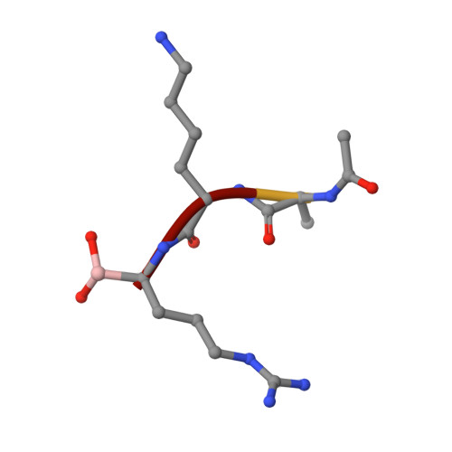
2.4 A Resolution Crystal Structure of the Prototypical Hormone-Processing Protease Kex2 in Complex with an Ala-Lys-Arg Boronic Acid Inhibitor

Holyoak, T.Wilson, M.A.Fenn, T.D.Kettner, C.A.Petsko, G.A.Fuller, R.S.Ringe, D.

(2003) Biochemistry 42: 6709-6718

  • DOI: https://doi.org/10.1021/bi034434t
  • Primary Citation of Related Structures:  
    1OT5

  • PubMed Abstract: 

    This paper reports the first structure of a member of the Kex2/furin family of eukaryotic pro-protein processing proteases, which cleave sites consisting of pairs or clusters of basic residues. Reported is the 2.4 A resolution crystal structure of the two-domain protein ssKex2 in complex with an Ac-Ala-Lys-boroArg inhibitor (R = 20.9%, R(free) = 24.5%). The Kex2 proteolytic domain is similar in its global fold to the subtilisin-like superfamily of degradative proteases. Analysis of the complex provides a structural basis for the extreme selectivity of this enzyme family that has evolved from a nonspecific subtilisin-like ancestor. The P-domain of ssKex2 has a novel jelly roll like fold consisting of nine beta strands and may potentially be involved, along with the buried Ca(2+) ion, in creating the highly determined binding site for P(1) arginine.


  • Organizational Affiliation

    Rosenstiel Basic Medical Sciences Research Center, Brandeis University, Waltham, Massachusetts 02454, USA.


Macromolecules
Find similar proteins by:  (by identity cutoff)  |  3D Structure
Entity ID: 1
MoleculeChains Sequence LengthOrganismDetailsImage
Kexin
A, B
477Saccharomyces cerevisiaeMutation(s): 0 
Gene Names: KEX2 OR QDS1 OR YNL238W OR N1122
EC: 3.4.21.61
UniProt
Find proteins for P13134 (Saccharomyces cerevisiae (strain ATCC 204508 / S288c))
Explore P13134 
Go to UniProtKB:  P13134
Entity Groups  
Sequence Clusters30% Identity50% Identity70% Identity90% Identity95% Identity100% Identity
UniProt GroupP13134
Glycosylation
Glycosylation Sites: 2Go to GlyGen: P13134-1
Sequence Annotations
Expand
  • Reference Sequence

Find similar proteins by:  Sequence   |   3D Structure  

Entity ID: 2
MoleculeChains Sequence LengthOrganismDetailsImage
Ac-Ala-Lys-boroArg N-acetylated boronic acid peptide inhibitor
C, D
4N/AMutation(s): 0 
Sequence Annotations
Expand
  • Reference Sequence
Oligosaccharides

Help

Entity ID: 3
MoleculeChains Length2D Diagram Glycosylation3D Interactions
2-acetamido-2-deoxy-beta-D-glucopyranose-(1-4)-2-acetamido-2-deoxy-beta-D-glucopyranose
E, F
2N-Glycosylation
Glycosylation Resources
GlyTouCan:  G42666HT
GlyCosmos:  G42666HT
GlyGen:  G42666HT
Small Molecules
Ligands 2 Unique
IDChains Name / Formula / InChI Key2D Diagram3D Interactions
NAG
Query on NAG

Download Ideal Coordinates CCD File 
G [auth A],
K [auth B]
2-acetamido-2-deoxy-beta-D-glucopyranose
C8 H15 N O6
OVRNDRQMDRJTHS-FMDGEEDCSA-N
CA
Query on CA

Download Ideal Coordinates CCD File 
H [auth A]
I [auth A]
J [auth A]
L [auth B]
M [auth B]
CALCIUM ION
Ca
BHPQYMZQTOCNFJ-UHFFFAOYSA-N
Modified Residues  1 Unique
IDChains TypeFormula2D DiagramParent
BOR
Query on BOR
C, D
PEPTIDE-LIKEC5 H15 B N4 O2ARG
Experimental Data & Validation

Experimental Data

  • Method: X-RAY DIFFRACTION
  • Resolution: 2.40 Å
  • R-Value Free:  0.245 (Depositor), 0.239 (DCC) 
  • R-Value Work:  0.209 (Depositor), 0.205 (DCC) 
  • R-Value Observed: 0.218 (Depositor) 
Space Group: P 65 2 2
Unit Cell:
Length ( Å )Angle ( ˚ )
a = 113.844α = 90
b = 113.844β = 90
c = 370.218γ = 120
Software Package:
Software NamePurpose
CNSrefinement
HKL-2000data reduction
SCALEPACKdata scaling
SOLVEphasing
RESOLVEphasing

Structure Validation

View Full Validation Report


Ligand Structure Quality Assessment 

Created with Raphaël 2.3.0Worse 01 BetterLigand structure goodness of fit to experimental dataBest fitted NAGClick on this verticalbar to view details

Entry History 

Deposition Data

Revision History  (Full details and data files)

  • Version 1.0: 2003-06-17
    Type: Initial release
  • Version 1.1: 2008-04-29
    Changes: Version format compliance
  • Version 1.2: 2011-07-13
    Changes: Atomic model, Database references, Derived calculations, Non-polymer description, Structure summary, Version format compliance
  • Version 1.3: 2012-12-12
    Changes: Other
  • Version 1.4: 2013-02-20
    Changes: Other
  • Version 1.5: 2016-12-14
    Changes: Structure summary
  • Version 2.0: 2020-07-29
    Type: Remediation
    Reason: Carbohydrate remediation
    Changes: Atomic model, Data collection, Derived calculations, Structure summary
  • Version 2.1: 2024-10-30
    Changes: Data collection, Database references, Structure summary