1PH8 | pdb_00001ph8

CRYSTAL STRUCTURE OF THE OXYTRICHA NOVA TELOMERE END-BINDING PROTEIN COMPLEXED WITH NONCOGNATE SSDNA GGGGTTTTGCGG


Experimental Data Snapshot

  • Method: X-RAY DIFFRACTION
  • Resolution: 2.36 Å
  • R-Value Free: 
    0.256 (Depositor), 0.256 (DCC) 
  • R-Value Work: 
    0.222 (Depositor), 0.222 (DCC) 
  • R-Value Observed: 
    0.222 (Depositor) 

wwPDB Validation 3D Report Full Report

Validation slider image for 1PH8

This is version 1.3 of the entry. See complete history

Literature

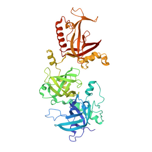
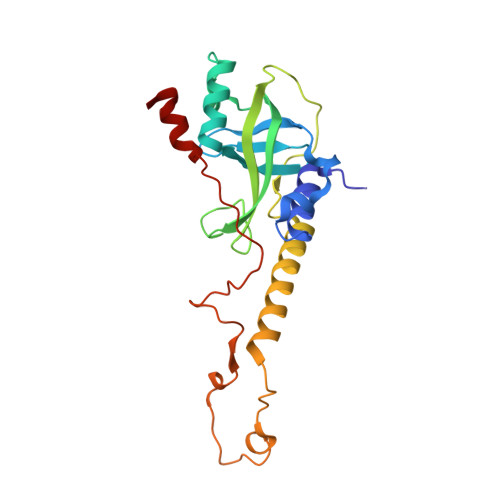
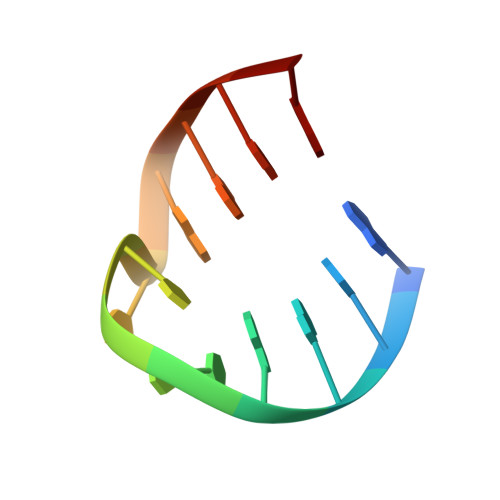
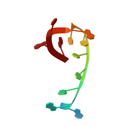
Nucleotide Shuffling and Ssdna Recognition in Oxytricha Nova Telomere End-Binding Protein Complexes

Theobald, D.L.Schultz, S.C.

(2003) EMBO J 22: 4314-4324

  • DOI: https://doi.org/10.1093/emboj/cdg415
  • Primary Citation Related Structures: 
    1PA6, 1PH1, 1PH2, 1PH3, 1PH4, 1PH5, 1PH6, 1PH7, 1PH8, 1PH9, 1PHJ

  • PubMed Abstract: 

    Sequence-specific protein recognition of single-stranded nucleic acids is critical for many fundamental cellular processes, such as DNA replication, DNA repair, transcription, translation, recombination, apoptosis and telomere maintenance. To explore the mechanisms of sequence-specific ssDNA recognition, we determined the crystal structures of 10 different non-cognate ssDNAs complexed with the Oxytricha nova telomere end-binding protein (OnTEBP) and evaluated their corresponding binding affinities (PDB ID codes 1PH1-1PH9 and 1PHJ). The thermodynamic and structural effects of these sequence perturbations could not have been predicted based solely upon the cognate structure. OnTEBP accommodates non-cognate nucleotides by both subtle adjustments and surprisingly large structural rearrangements in the ssDNA. In two complexes containing ssDNA intermediates that occur during telomere extension by telomerase, entire nucleotides are expelled from the complex. Concurrently, the sequence register of the ssDNA shifts to re-establish a more cognate-like pattern. This phenomenon, termed nucleotide shuffling, may be of general importance in protein recognition of single-stranded nucleic acids. This set of structural and thermodynamic data highlights a fundamental difference between protein recognition of ssDNA versus dsDNA.


  • Organizational Affiliation
    • Department of Chemistry and Biochemistry, University of Colorado at Boulder, Boulder, CO 80309-0215, USA. theobal@colorado.edu

Macromolecule Content 

  • Total Structure Weight: 89.06 kDa 
  • Atom Count: 6,681 
  • Modeled Residue Count: 711 
  • Deposited Residue Count: 714 
  • Unique protein chains: 2
  • Unique nucleic acid chains: 2

Macromolecules


Find similar proteins by:|  3D Structure
Entity ID: 3
MoleculeChains  Sequence LengthOrganismDetailsImage
Telomere-binding protein alpha subunitD [auth A]460Sterkiella novaMutation(s): 0 
Gene Names: MAC-56A AND MAC-56K AND MAC-56S
UniProt
Find proteins for P29549 (Sterkiella nova)
Explore P29549 
Go to UniProtKB:  P29549
Entity Groups
Sequence Clusters30% Identity50% Identity70% Identity90% Identity95% Identity100% Identity
UniProt GroupP29549
Sequence Annotations
Expand
Reference Sequence
Find similar proteins by:|  3D Structure
Entity ID: 4
MoleculeChains  Sequence LengthOrganismDetailsImage
Telomere-binding protein beta subunitE [auth B]216Sterkiella novaMutation(s): 0 
Gene Names: MAC-41A AND MAC-41S
UniProt
Find proteins for P16458 (Sterkiella nova)
Explore P16458 
Go to UniProtKB:  P16458
Entity Groups
Sequence Clusters30% Identity50% Identity70% Identity90% Identity95% Identity100% Identity
UniProt GroupP16458
Sequence Annotations
Expand
Reference Sequence
Find similar nucleic acids by:  Sequence
Entity ID: 1
MoleculeChains LengthOrganismImage
5'-D(*GP*GP*GP*GP*TP*TP*TP*TP*GP*GP*GP*GP*T)-3'A [auth G],
C [auth H]
13N/A
Sequence Annotations
Expand
Reference Sequence
Find similar nucleic acids by:  Sequence
Entity ID: 2
MoleculeChains LengthOrganismImage
5'-D(*GP*GP*GP*GP*TP*TP*TP*TP*GP*CP*GP*G)-3'B [auth D]12N/A
Sequence Annotations
Expand
Reference Sequence

Experimental Data & Validation

Experimental Data

  • Method: X-RAY DIFFRACTION
  • Resolution: 2.36 Å
  • R-Value Free:  0.256 (Depositor), 0.256 (DCC) 
  • R-Value Work:  0.222 (Depositor), 0.222 (DCC) 
  • R-Value Observed: 0.222 (Depositor) 
Space Group: P 61 2 2
Unit Cell:
Length ( Å )Angle ( ˚ )
a = 93.472α = 90
b = 93.472β = 90
c = 423.554γ = 120
Software Package:
Software NamePurpose
DENZOdata reduction
SCALEPACKdata scaling
CNSrefinement
CNSphasing

Structure Validation

View Full Validation Report


Entry History 

Deposition Data

Revision History  (Full details and data files)

  • Version 1.0: 2003-06-17
    Type: Initial release
  • Version 1.1: 2008-04-29
    Changes: Version format compliance
  • Version 1.2: 2011-07-13
    Changes: Version format compliance
  • Version 1.3: 2024-02-14
    Changes: Data collection, Database references, Derived calculations