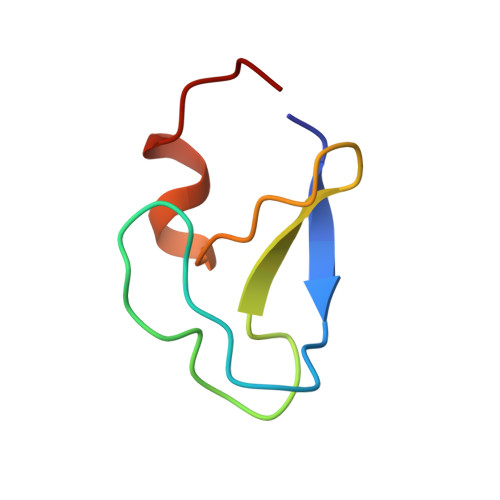
Crystal structure of the cys2 activator-binding domain of protein kinase C delta in complex with phorbol ester.
Zhang, G., Kazanietz, M.G., Blumberg, P.M., Hurley, J.H.(1995) Cell 81: 917-924
- PubMed: 7781068
- DOI: https://doi.org/10.1016/0092-8674(95)90011-x
- Primary Citation of Related Structures:
1PTQ, 1PTR - PubMed Abstract:
Protein kinase Cs (PKCs) are a ubiquitous family of regulatory enzymes that associate with membranes and are activated by diacylglycerol or tumor-promoting agonists such as phorbol esters. The structure of the second activator-binding domain of PKC delta has been determined in complex with phorbol 13-acetate, which binds in a groove between two pulled-apart beta strands at the tip of the domain. The C3, C4, and C20 phorbol oxygens form hydrogen bonds with main-chain groups whose orientation is controlled by a set of highly conserved residues. Phorbol binding caps the groove and forms a contiguous hydrophobic surface covering one-third of the domain, explaining how the activator promotes insertion of PKC into membranes.
- Laboratory of Molecular Biology, National Institute of Diabetes and Digestive and Kidney Diseases, National Institutes of Health, Bethesda, Maryland 20892-0580, USA.
Organizational Affiliation:


