1Q3I | pdb_00001q3i

Crystal Structure of Na,K-ATPase N-domain


Experimental Data Snapshot

  • Method: X-RAY DIFFRACTION
  • Resolution: 2.60 Å
  • R-Value Free: 
    0.292 (Depositor), 0.253 (DCC) 
  • R-Value Work: 
    0.251 (Depositor), 0.254 (DCC) 

wwPDB Validation 3D Report Full Report

Validation slider image for 1Q3I

This is version 1.5 of the entry. See complete history

Literature

The Crystallographic Structure of Na,K-ATPase N-domain at 2.6 A Resolution

Hakansson, K.O.

(2003) J Mol Biology 332: 1175-1182

  • DOI: https://doi.org/10.1016/j.jmb.2003.07.012
  • Primary Citation Related Structures: 
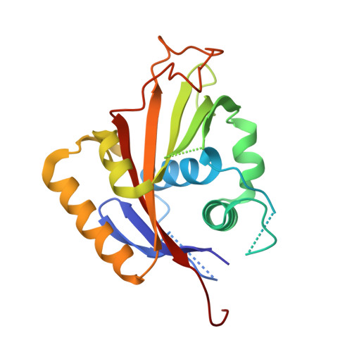
    1Q3I

  • PubMed Abstract: 

    The structure of the N-domain of porcine alpha(2) Na,K-ATPase was determined crystallographically to 3.2A resolution by isomorphous heavy-atom replacement using a single mercury derivative. The structure was finally refined against 2.6A resolution synchrotron data. The domain forms a seven-stranded antiparallel beta-sheet with two additional beta-strands forming a hairpin and five alpha-helices. Approximately 75% of the residues were superimposable with residues from the structure of Ca-ATPase N-domain, and a structure-based sequence alignment is presented. The positions of key residues are discussed in relation to the pattern of hydrophobicity, charge and sequence conservation of the molecular surface. The structure of a hexahistidine tag binding to nickel ions is presented.


  • Organizational Affiliation
    • August Krogh Institute, Copenhagen University, Universitetsparken 13, DK-2100 OE, Copenhagen, Denmark. kohakansson@aki.ku.dk

Macromolecule Content 

  • Total Structure Weight: 24.3 kDa 
  • Atom Count: 1,612 
  • Modeled Residue Count: 192 
  • Deposited Residue Count: 214 
  • Unique protein chains: 1

Macromolecules

Find similar proteins by:|  3D Structure
Entity ID: 1
MoleculeChains  Sequence LengthOrganismDetailsImage
Na,K-ATPase214Sus scrofaMutation(s): 0 
EC: 3.6.3.9 (PDB Primary Data), 7.2.2.13 (UniProt)
UniProt
Find proteins for D2WKD8 (Sus scrofa)
Explore D2WKD8 
Go to UniProtKB:  D2WKD8
Entity Groups
Sequence Clusters30% Identity50% Identity70% Identity90% Identity95% Identity100% Identity
UniProt GroupD2WKD8
Sequence Annotations
Expand
Reference Sequence

Experimental Data & Validation

Experimental Data

  • Method: X-RAY DIFFRACTION
  • Resolution: 2.60 Å
  • R-Value Free:  0.292 (Depositor), 0.253 (DCC) 
  • R-Value Work:  0.251 (Depositor), 0.254 (DCC) 
Space Group: F 2 3
Unit Cell:
Length ( Å )Angle ( ˚ )
a = 147.31α = 90
b = 147.31β = 90
c = 147.31γ = 90
Software Package:
Software NamePurpose
MOSFLMdata reduction
SCALAdata scaling
CNSrefinement
CCP4data scaling
CNSphasing

Structure Validation

View Full Validation Report


Entry History 

Deposition Data

Revision History  (Full details and data files)

  • Version 1.0: 2004-08-03
    Type: Initial release
  • Version 1.1: 2008-04-29
    Changes: Version format compliance
  • Version 1.2: 2011-07-13
    Changes: Derived calculations, Version format compliance
  • Version 1.3: 2011-11-16
    Changes: Atomic model
  • Version 1.4: 2018-03-07
    Changes: Data collection
  • Version 1.5: 2024-02-14
    Changes: Data collection, Database references, Derived calculations