Restricted motion of the lipoyl-lysine swinging arm in the pyruvate dehydrogenase complex of Escherichia coli.
Jones, D.D., Stott, K.M., Howard, M.J., Perham, R.N.(2000) Biochemistry 39: 8448-8459
- PubMed: 10913250 Search on PubMed
- DOI: https://doi.org/10.1021/bi992978i
- Primary Citation Related Structures:
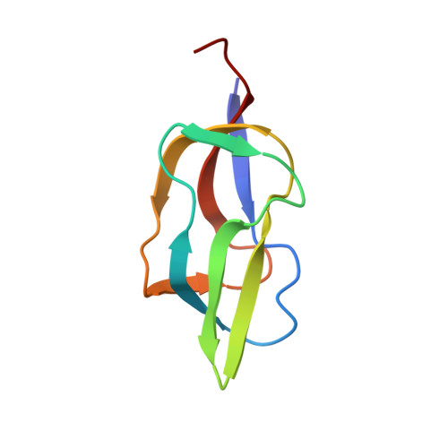
1QJO - PubMed Abstract:
The three lipoyl (E2plip) domains of the dihydrolipoyl acetyltransferase component of the pyruvate dehydrogenase (PDH) complex of Escherichia coli house the lipoyl-lysine side chain essential for active-site coupling and substrate channelling within the complex. The structure of the unlipoylated form of the innermost domain (E2plip(apo)) was determined by multidimensional NMR spectroscopy and found to resemble closely that of a nonfunctional hybrid domain determined previously [Green et al. (1995) J. Mol. Biol. 248, 328-343]. The domain comprises two four-stranded beta-sheets, with the target lysine residue residing at the tip of a type-I beta-turn in one of the sheets; the N- and C-termini lie close together at the opposite end of the molecule in the other beta-sheet. Measurement of (15)N NMR relaxation parameters and backbone hydrogen/deuterium (H/D) exchange rates reveals that the residues in and surrounding the lipoyl-lysine beta-turn in the E2plip(apo) form of the domain become less flexible after lipoylation of the lysine residue. This implies that the lipoyl-lysine side chain may not sample the full range of conformational space once thought. Moreover, reductive acetylation of the lipoylated domain (E2plip(holo) --> E2plip(redac)) was accompanied by large changes in chemical shift between the two forms, and multiple resonances were observed for several residues. This implies a change in conformation and the existence of multiple conformations of the domain on reductive acetylation, which may be important in stabilizing this catalytic intermediate.
- Cambridge Centre for Molecular Recognition, Department of Biochemistry, University of Cambridge, UK.
Organizational Affiliation: