1QOP

CRYSTAL STRUCTURE OF WILD-TYPE TRYPTOPHAN SYNTHASE COMPLEXED WITH INDOLE PROPANOL PHOSPHATE


Experimental Data Snapshot

  • Method: X-RAY DIFFRACTION
  • Resolution: 1.40 Å
  • R-Value Free: 
    0.177 (Depositor), 0.180 (DCC) 
  • R-Value Work: 
    0.150 (Depositor), 0.150 (DCC) 

Starting Model: experimental
View more details

wwPDB Validation   3D Report Full Report


Ligand Structure Quality Assessment 

Created with Raphaël 2.3.0Worse 01 BetterLigand structure goodness of fit to experimental dataBest fitted IPLClick on this verticalbar to view detailsBest fitted PLPClick on this verticalbar to view details

This is version 1.4 of the entry. See complete history


Literature

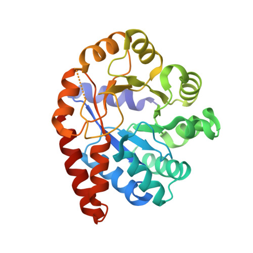
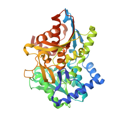
Crystal Structure of Wild-Type Tryptophan Synthase Complexed with the Natural Substrate Indole-3-Glycerol Phosphate

Weyand, M.Schlichting, I.

(1999) Biochemistry 38: 16469

  • DOI: https://doi.org/10.1021/bi9920533
  • Primary Citation of Related Structures:  
    1QOP, 1QOQ

  • PubMed Abstract: 

    We used freeze trapping to stabilize the Michaelis complex of wild-type tryptophan synthase and the alpha-subunit substrate indole-3-glycerol phosphate (IGP) and determined its structure to 1. 8 A resolution. In addition, we determined the 1.4 A resolution structure of the complex with indole-3-propanole phosphate (IPP), a noncleavable IGP analogue. The interaction of the 3'-hydroxyl of IGP with the catalytic alphaGlu49 leads to a twisting of the propane chain and to a repositioning of the indole ring compared to IPP. Concomitantly, the catalytic alphaAsp60 rotates resulting in a translocation of the COMM domain [betaGly102-betaGly189, for definition see Schneider et al. (1998) Biochemistry 37, 5394-5406] in a direction opposite to the one in the IPP complex. This results in loss of the allosteric sodium ion bound at the beta-subunit and an opening of the beta-active site, thereby making the cofactor pyridoxal 5'-phosphate (PLP) accessible to solvent and thus serine binding. These findings form the structural basis for the information transfer from the alpha- to the beta-subunit and may explain the affinity increase of the beta-active site for serine upon IGP binding.


  • Organizational Affiliation

    Max-Planck-Institute for Molecular Physiology, Department of Physical Biochemistry, Dortmund, Germany.


Macromolecules
Find similar proteins by:  (by identity cutoff)  |  3D Structure
Entity ID: 1
MoleculeChains Sequence LengthOrganismDetailsImage
TRYPTOPHAN SYNTHASE ALPHA CHAIN268Salmonella enterica subsp. enterica serovar TyphimuriumMutation(s): 0 
Gene Names: TRPA
EC: 4.2.1.20
UniProt
Find proteins for P00929 (Salmonella typhimurium (strain LT2 / SGSC1412 / ATCC 700720))
Explore P00929 
Go to UniProtKB:  P00929
Entity Groups  
Sequence Clusters30% Identity50% Identity70% Identity90% Identity95% Identity100% Identity
UniProt GroupP00929
Sequence Annotations
Expand
  • Reference Sequence
Find similar proteins by:  (by identity cutoff)  |  3D Structure
Entity ID: 2
MoleculeChains Sequence LengthOrganismDetailsImage
TRYPTOPHAN SYNTHASE BETA CHAIN396Salmonella enterica subsp. enterica serovar TyphimuriumMutation(s): 0 
Gene Names: TRPB
EC: 4.2.1.20
UniProt
Find proteins for P0A2K1 (Salmonella typhimurium (strain LT2 / SGSC1412 / ATCC 700720))
Explore P0A2K1 
Go to UniProtKB:  P0A2K1
Entity Groups  
Sequence Clusters30% Identity50% Identity70% Identity90% Identity95% Identity100% Identity
UniProt GroupP0A2K1
Sequence Annotations
Expand
  • Reference Sequence
Small Molecules
Binding Affinity Annotations 
IDSourceBinding Affinity
IPL BindingDB:  1QOP Ki: 1.50e+4 (nM) from 1 assay(s)
Experimental Data & Validation

Experimental Data

  • Method: X-RAY DIFFRACTION
  • Resolution: 1.40 Å
  • R-Value Free:  0.177 (Depositor), 0.180 (DCC) 
  • R-Value Work:  0.150 (Depositor), 0.150 (DCC) 
Space Group: C 1 2 1
Unit Cell:
Length ( Å )Angle ( ˚ )
a = 182.2α = 90
b = 60.3β = 94.7
c = 67.4γ = 90
Software Package:
Software NamePurpose
REFMACrefinement
XDSdata reduction
XSCALEdata scaling
CNSphasing

Structure Validation

View Full Validation Report


Ligand Structure Quality Assessment 

Created with Raphaël 2.3.0Worse 01 BetterLigand structure goodness of fit to experimental dataBest fitted IPLClick on this verticalbar to view detailsBest fitted PLPClick on this verticalbar to view details

Entry History 

Deposition Data

Revision History  (Full details and data files)

  • Version 1.0: 2000-11-10
    Type: Initial release
  • Version 1.1: 2013-04-17
    Changes: Data collection, Derived calculations, Non-polymer description, Other, Source and taxonomy, Version format compliance
  • Version 1.2: 2019-05-08
    Changes: Data collection, Derived calculations, Experimental preparation, Other
  • Version 1.3: 2019-07-24
    Changes: Data collection
  • Version 1.4: 2023-12-13
    Changes: Data collection, Database references, Derived calculations, Other, Refinement description