Hot-spot mimicry of a cytokine receptor by a small molecule.
Thanos, C.D., DeLano, W.L., Wells, J.A.(2006) Proc Natl Acad Sci U S A 103: 15422-15427
- PubMed: 17032757 Search on PubMedSearch on PubMed Central
- DOI: https://doi.org/10.1073/pnas.0607058103
- Primary Citation Related Structures:
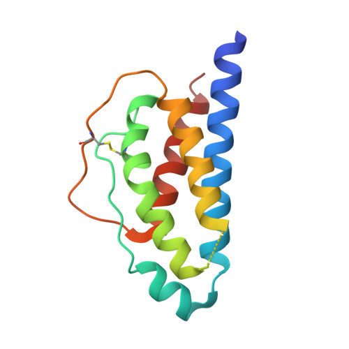
1QVN - PubMed Abstract:
Protein-protein complexes remain enticing, but extremely challenging, targets for small-molecule drug discovery. In a rare example described earlier, a high-affinity small molecule, SP4206 (Kd approximately 70 nM), was found to block binding of the IL-2alpha receptor (IL-2Ralpha) to IL-2 (Kd approximately 10 nM). Recently, the structure of the IL-2/IL-2Ralpha complex was solved [Rickert, M., Wang, X., Boulanger, M. J., Goriatcheva, N., Garcia, K. C. (2005) Science 308:1477-1480]. Using structural and functional analysis, we compare how SP4206 mimics the 83-fold larger IL-2Ralpha in binding IL-2. The binding free energy per contact atom (ligand efficiency) for SP4206 is about twice that of the receptor because of a smaller, but overlapping, contact epitope that insinuates into grooves and cavities not accessed by the receptor. Despite its independent design, the small molecule has a similar, but more localized, charge distribution compared with IL-2Ralpha. Mutational studies show that SP4206 targets virtually the same critical "hot-spot" residues on IL-2 that drive binding of IL-2Ralpha. Moreover, a mutation that enhances binding to the IL-2Ralpha near these hot spots also enhances binding to SP4206. Although the protein and small molecule do bind the same hot spot, they trap very different conformations of IL-2 because of its flexible nature. Our studies suggest that precise structural mimics of receptors are not required for high-affinity binding of small molecules, and they show that there are multiple solutions to tight binding at shared and adaptive hot spots.
- Sunesis Pharmaceuticals, 341 Oyster Point Boulevard, South San Francisco, CA 94080, USA.
Organizational Affiliation: