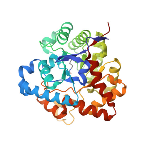
1QW7

Structure of an Engineered Organophosphorous Hydrolase with Increased Activity Toward Hydrolysis of Phosphothiolate Bonds


Experimental Data Snapshot

  • Method: X-RAY DIFFRACTION
  • Resolution: 1.90 Å
  • R-Value Free: 0.230 
  • R-Value Work: 0.179 
  • R-Value Observed: 0.179 

Starting Model: experimental
View more details

wwPDB Validation   3D Report Full Report


Ligand Structure Quality Assessment 


This is version 1.5 of the entry. See complete history


Literature

Structural and mutational studies of organophosphorus hydrolase reveal a cryptic and functional allosteric-binding site.

Grimsley, J.K.Calamini, B.Wild, J.R.Mesecar, A.D.

(2005) Arch Biochem Biophys 442: 169-179

  • DOI: https://doi.org/10.1016/j.abb.2005.08.012
  • Primary Citation of Related Structures:  
    1QW7

  • PubMed Abstract: 

    Organophosphorus hydrolase detoxifies a broad range of organophosphate pesticides and the chemical warfare agents (CWAs) sarin and VX. Previously, rational genetic engineering produced OPH variants with 30-fold enhancements in the hydrolysis of CWA and their analogs. One interesting variant (H254R) in which the histidine at position 254 was changed to an arginine showed a 4-fold increase in the hydrolysis of demetonS (VX analog), a 14-fold decrease with paraoxon (an insecticide), and a 183-fold decrease with DFP (sarin analog). The three-dimensional structure of this enzyme at 1.9A resolution with the inhibitor, diethyl 4-methylbenzylphosphonate (EBP), revealed that the inhibitor did not bind at the active site, but bound exclusively into a well-defined surface pocket 12 A away from the active site. This structural feature was accompanied by non-competitive inhibition of paraoxon hydrolysis by EBP with H254R, in contrast to the native enzyme, which showed competitive inhibition. These parallel structure-function characteristics identify a functional, allosteric site on the surface of this enzyme.


  • Organizational Affiliation

    Biochemistry and Biophysics Department, Texas A&M University, College Station, TX 77843-2128, USA.


Macromolecules
Find similar proteins by:  (by identity cutoff)  |  3D Structure
Entity ID: 1
MoleculeChains Sequence LengthOrganismDetailsImage
Parathion hydrolase
A, B
336Brevundimonas diminutaMutation(s): 1 
Gene Names: OPD
EC: 3.1.8.1
UniProt
Find proteins for P0A434 (Brevundimonas diminuta)
Go to UniProtKB:  P0A434
Sequence Annotations
Expand
  • Reference Sequence
Small Molecules
Modified Residues  1 Unique
IDChains TypeFormula2D DiagramParent
KCX
Query on KCX
A, B
L-PEPTIDE LINKINGC7 H14 N2 O4LYS
Binding Affinity Annotations 
IDSourceBinding Affinity
EBP PDBBind:  1QW7 Ki: 2.50e+5 (nM) from 1 assay(s)
Experimental Data & Validation

Experimental Data

  • Method: X-RAY DIFFRACTION
  • Resolution: 1.90 Å
  • R-Value Free: 0.230 
  • R-Value Work: 0.179 
  • R-Value Observed: 0.179 
  • Space Group: C 1 2 1
Unit Cell:
Length ( Å )Angle ( ˚ )
a = 126.719α = 90
b = 89.879β = 91.73
c = 68.337γ = 90
Software Package:
Software NamePurpose
CNSrefinement
DENZOdata reduction
SCALEPACKdata scaling
CNSphasing

Structure Validation

View Full Validation Report


Ligand Structure Quality Assessment 


Entry History 

Deposition Data

Revision History  (Full details and data files)

  • Version 1.0: 2004-11-30
    Type: Initial release
  • Version 1.1: 2008-04-29
    Changes: Version format compliance
  • Version 1.2: 2011-07-13
    Changes: Version format compliance
  • Version 1.3: 2021-10-27
    Changes: Database references, Derived calculations
  • Version 1.4: 2023-08-16
    Changes: Data collection, Refinement description
  • Version 1.5: 2023-11-15
    Changes: Data collection