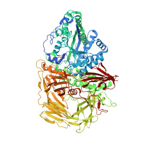
Insights into the mechanism of Drosophila melanogaster Golgi alpha-mannosidase II through the structural analysis of covalent reaction intermediates.
Numao, S., Kuntz, D.A., Withers, S.G., Rose, D.R.(2003) J Biol Chem 278: 48074-48083
- PubMed: 12960159
- DOI: https://doi.org/10.1074/jbc.M309249200
- Primary Citation of Related Structures:
1QWN, 1QWU, 1QX1 - PubMed Abstract:
The family 38 golgi alpha-mannosidase II, thought to cleave mannosidic bonds through a double displacement mechanism involving a reaction intermediate, is a clinically important enzyme involved in glycoprotein processing. The structure of three different covalent glycosyl-enzyme intermediates have been determined to 1.2-A resolution for the Golgi alpha-mannosidase II from Drosophila melanogaster by use of fluorinated sugar analogues, both with the wild-type enzyme and a mutant enzyme in which the acid/base catalyst has been removed. All these structures reveal sugar intermediates bound in a distorted 1S5 skew boat conformation. The similarity of this conformation with that of the substrate in the recently determined structure of the Michaelis complex of a beta-mannanase (Ducros, V. M. A., Zechel, D. L., Murshudov, G. N., Gilbert, H. J., Szabo, L., Stoll, D., Withers, S. G., and Davies, G. J. (2002) Angew. Chem. Int. Ed. Engl. 41, 2824-2827) suggests that these disparate enzymes have recruited common stereoelectronic features in evolving their catalytic mechanisms.
Organizational Affiliation:
Department of Chemistry, University of British Columbia, Vancouver, British Columbia V6T 1Z1, Canada.


