1RT1 | pdb_00001rt1

CRYSTAL STRUCTURE OF HIV-1 REVERSE TRANSCRIPTASE COMPLEXED WITH MKC-442


Experimental Data Snapshot

  • Method: X-RAY DIFFRACTION
  • Resolution: 2.55 Å
  • R-Value Work: 
    0.197 (Depositor) 
  • R-Value Observed: 
    0.197 (Depositor) 

Starting Models: experimental
View more details

wwPDB Validation 3D Report Full Report

Validation slider image for 1RT1

This is version 1.4 of the entry. See complete history

Literature

Complexes of HIV-1 reverse transcriptase with inhibitors of the HEPT series reveal conformational changes relevant to the design of potent non-nucleoside inhibitors.

Hopkins, A.L.Ren, J.Esnouf, R.M.Willcox, B.E.Jones, E.Y.Ross, C.Miyasaka, T.Walker, R.T.Tanaka, H.Stammers, D.K.Stuart, D.I.

(1996) J Med Chem 39: 1589-1600

  • DOI: https://doi.org/10.1021/jm960056x
  • Primary Citation Related Structures: 
    1RT1, 1RT2

  • PubMed Abstract: 

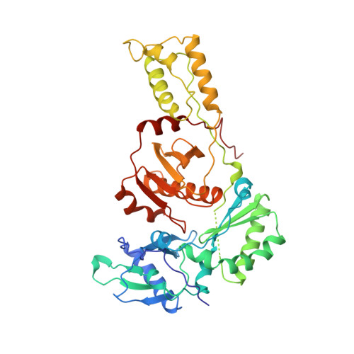
    Crystal structures of HIV-1 reverse transcriptase (RT) complexed with a range of chemically diverse non-nucleoside inhibitors (NNIs) have shown a single pocket in which the inhibitors bind and details of the inhibitor-protein interactions. To delineate the structural requirements for an effective inhibitor, we have determined the structures of three closely related NNIs which vary widely in their potencies. Crystal structures of HIV-1 RT complexed with two very potent inhibitors, MKC-442 and TNK-651, at 2.55 angstroms resolution complement our previous analysis of the complex with the less effective inhibitor, HEPT. These structures reveal conformational changes which correlate with changes in potency. We suggest that a major determinant of increased potency in the analogues of HEPT is an improved interaction between residue Tyr181 in the protein and the 6-benzyl ring of the inhibitors which stabilizes the structure of the complex. This arises through a conformational switching of the protein structure triggered by the steric bulk of the 5-substituent of the inhibitor pyrimidine ring.


  • Organizational Affiliation
    • Laboratory of Molecular Biophysics, Rex Richards Building, Oxford, UK.

Macromolecule Content 

  • Total Structure Weight: 116.3 kDa 
  • Atom Count: 8,272 
  • Modeled Residue Count: 954 
  • Deposited Residue Count: 1,000 
  • Unique protein chains: 2

Macromolecules

Find similar proteins by:|  3D Structure
Entity ID: 1
MoleculeChains  Sequence LengthOrganismDetailsImage
HIV-1 REVERSE TRANSCRIPTASE560Human immunodeficiency virus 1Mutation(s): 1 
Gene Names: HIV-1 POL
EC: 2.7.7.49
UniProt
Find proteins for P04585 (Human immunodeficiency virus type 1 group M subtype B (isolate HXB2))
Explore P04585 
Go to UniProtKB:  P04585
Entity Groups
Sequence Clusters30% Identity50% Identity70% Identity90% Identity95% Identity100% Identity
UniProt GroupP04585
Sequence Annotations
Expand
Reference Sequence
Find similar proteins by:|  3D Structure
Entity ID: 2
MoleculeChains  Sequence LengthOrganismDetailsImage
HIV-1 REVERSE TRANSCRIPTASE440Human immunodeficiency virus 1Mutation(s): 0 
Gene Names: HIV-1 POL
EC: 2.7.7.49
UniProt
Find proteins for P04585 (Human immunodeficiency virus type 1 group M subtype B (isolate HXB2))
Explore P04585 
Go to UniProtKB:  P04585
Entity Groups
Sequence Clusters30% Identity50% Identity70% Identity90% Identity95% Identity100% Identity
UniProt GroupP04585
Sequence Annotations
Expand
Reference Sequence

Small Molecules

Ligands 1 Unique
IDChains Name / Formula / InChI Key2D Diagram3D Interactions
MKC

Query on MKC


Download:Ideal Coordinates CCD File
C [auth A]6-BENZYL-1-ETHOXYMETHYL-5-ISOPROPYL URACIL
C17 H22 N2 O3
MLILORUFDVLTSP-UHFFFAOYSA-N
Modified Residues  1 Unique
IDChains TypeFormula2D DiagramParent
CSD
Query on CSD
A
L-PEPTIDE LINKINGC3 H7 N O4 SCYS
Binding Affinity Annotations 
IDSourceBinding Affinity
MKC BindingDB:  1RT1 IC50: min: 8, max: 800 (nM) from 5 assay(s)
EC50: min: 2, max: 20 (nM) from 4 assay(s)

Experimental Data & Validation

Experimental Data

  • Method: X-RAY DIFFRACTION
  • Resolution: 2.55 Å
  • R-Value Work:  0.197 (Depositor) 
  • R-Value Observed: 0.197 (Depositor) 
Space Group: P 21 21 21
Unit Cell:
Length ( Å )Angle ( ˚ )
a = 136.8α = 90
b = 109.8β = 90
c = 72.4γ = 90
Software Package:
Software NamePurpose
X-PLORmodel building
X-PLORrefinement
DENZOdata reduction
SCALEPACKdata scaling
X-PLORphasing

Structure Validation

View Full Validation Report


Entry History 

Deposition Data

Revision History  (Full details and data files)

  • Version 1.0: 1997-04-21
    Type: Initial release
  • Version 1.1: 2008-03-24
    Changes: Version format compliance
  • Version 1.2: 2011-07-13
    Changes: Version format compliance
  • Version 1.3: 2023-08-09
    Changes: Database references, Derived calculations, Other, Refinement description
  • Version 1.4: 2024-11-13
    Changes: Data collection, Structure summary