1SAE | pdb_00001sae

HIGH RESOLUTION SOLUTION NMR STRUCTURE OF THE OLIGOMERIZATION DOMAIN OF P53 BY MULTI-DIMENSIONAL NMR (SAC STRUCTURES)


Experimental Data Snapshot

  • Method: SOLUTION NMR
  • Conformers Submitted: 77 

wwPDB Validation 3D Report Full Report

Validation slider image for 1SAE

This is version 1.4 of the entry. See complete history

Literature

Refined solution structure of the oligomerization domain of the tumour suppressor p53.

Clore, G.M.Ernst, J.Clubb, R.Omichinski, J.G.Kennedy, W.M.Sakaguchi, K.Appella, E.Gronenborn, A.M.

(1995) Nat Struct Biol 2: 321-333

  • DOI: https://doi.org/10.1038/nsb0495-321
  • Primary Citation Related Structures: 
    1SAE, 1SAF, 1SAK, 1SAL

  • PubMed Abstract: 

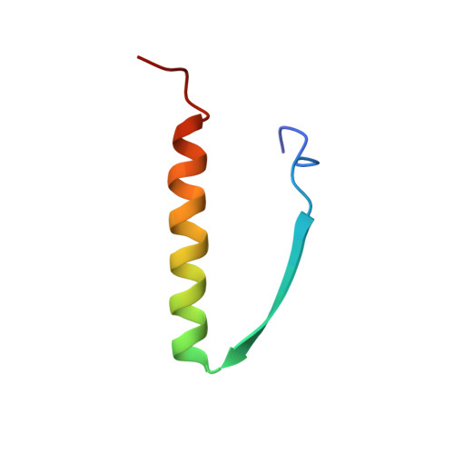
    The NMR solution structure of the oligomerization domain of the tumour suppressor p53 (residues 319-360) has been refined. The structure comprises a dimer of dimers, oriented in an approximately orthogonal manner. The present structure determination is based on 4,472 experimental NMR restraints which represents a three and half fold increase over our previous work in the number of NOE restraints at the tetramerization interface. A comparison with the recently solved 1.7 A resolution X-ray structure shows that the structures are very similar and that the average angular root-mean-square difference in the interhelical angles is about 1 degree. The results of recent extensive mutagenesis data and the possible effects of mutations which have been identified in human cancers are discussed in the light of the present structure.


  • Organizational Affiliation
    • Laboratory of Chemical Physics, National Institute of Diabetes and Digestive and Kidney Diseases, National Institutes of Health, Bethesda, MD 20892-0520, USA.

Macromolecule Content 

  • Total Structure Weight: 19.79 kDa 
  • Atom Count: 1,396 
  • Modeled Residue Count: 168 
  • Deposited Residue Count: 168 
  • Unique protein chains: 1

Macromolecules

Find similar proteins by:|  3D Structure
Entity ID: 1
MoleculeChains  Sequence LengthOrganismDetailsImage
TUMOR SUPPRESSOR P53
A, B, C, D
42Homo sapiensMutation(s): 0 
UniProt & NIH Common Fund Data Resources
Find proteins for P04637 (Homo sapiens)
Explore P04637 
Go to UniProtKB:  P04637
PHAROS:  P04637
GTEx:  ENSG00000141510 
Entity Groups
Sequence Clusters30% Identity50% Identity70% Identity90% Identity95% Identity100% Identity
UniProt GroupP04637
Sequence Annotations
Expand
Reference Sequence

Experimental Data & Validation

Experimental Data

  • Method: SOLUTION NMR
  • Conformers Submitted: 77 

Structure Validation

View Full Validation Report


Entry History 

Deposition Data

Revision History  (Full details and data files)

  • Version 1.0: 1995-10-15
    Type: Initial release
  • Version 1.1: 2008-03-24
    Changes: Version format compliance
  • Version 1.2: 2011-07-13
    Changes: Version format compliance
  • Version 1.3: 2022-03-02
    Changes: Database references, Derived calculations, Other
  • Version 1.4: 2024-05-22
    Changes: Data collection