1SB3 | pdb_00001sb3

Structure of 4-hydroxybenzoyl-CoA reductase from Thauera aromatica


Experimental Data Snapshot

  • Method: X-RAY DIFFRACTION
  • Resolution: 2.20 Å
  • R-Value Free: 
    0.205 (Depositor), 0.198 (DCC) 
  • R-Value Work: 
    0.171 (Depositor), 0.166 (DCC) 
  • R-Value Observed: 
    0.171 (Depositor) 

Starting Model: experimental
View more details

wwPDB Validation 3D Report Full Report

Validation slider image for 1SB3

Ligand Structure Quality Assessment 


This is version 1.3 of the entry. See complete history

Literature

Structure of a Xanthine Oxidase-Related 4-Hydroxybenzoyl-CoA Reductase with an Additional [4Fe-4S] Cluster and an Inverted Electron Flow.

Unciuleac, M.Warkentin, E.Page, C.C.Boll, M.Ermler, U.

(2004) Structure 12: 2249-2256

  • DOI: https://doi.org/10.1016/j.str.2004.10.008
  • Primary Citation Related Structures: 
    1RM6, 1SB3

  • PubMed Abstract: 

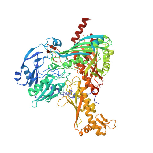
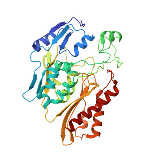
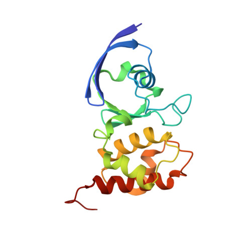
    The Mo-flavo-Fe/S-dependent heterohexameric protein complex 4-hydroxybenzoyl-CoA reductase (4-HBCR, dehydroxylating) is a central enzyme of the anaerobic degradation of phenolic compounds and belongs to the xanthine oxidase (XO) family of molybdenum enzymes. Its X-ray structure was established at 1.6 A resolution. The most pronounced difference between 4-HBCR and other structurally characterized members of the XO family is the insertion of 40 amino acids within the beta subunit, which carries an additional [4Fe-4S] cluster at a distance of 16.5 A to the isoalloxazine ring of FAD. The architecture of 4-HBCR and concomitantly performed electron transfer rate calculations suggest an inverted electron transfer chain from the donor ferredoxin via the [4Fe-4S] cluster to the Mo over a distance of 55 A. The binding site of 4-hydroxybenzoyl-CoA is located in an 18 A long channel lined up by several aromatic side chains around the aromatic moiety, which are proposed to shield and stabilize the postulated radical intermediates during catalysis.


  • Organizational Affiliation
    • Institut für Biologie II, Mikrobiologie Schänzlestrasse 1, D-79104 Freiburg, Germany.

Macromolecule Content 

  • Total Structure Weight: 273.1 kDa 
  • Atom Count: 19,897 
  • Modeled Residue Count: 2,485 
  • Deposited Residue Count: 2,508 
  • Unique protein chains: 3

Macromolecules

Find similar proteins by:|  3D Structure
Entity ID: 1
MoleculeChains  Sequence LengthOrganismDetailsImage
4-hydroxybenzoyl-CoA reductase alpha subunit
A, D
769Thauera aromaticaMutation(s): 0 
EC: 1.3.99.20 (PDB Primary Data), 1.1.7.1 (UniProt)
UniProt
Find proteins for O33819 (Thauera aromatica)
Explore O33819 
Go to UniProtKB:  O33819
Entity Groups
Sequence Clusters30% Identity50% Identity70% Identity90% Identity95% Identity100% Identity
UniProt GroupO33819
Sequence Annotations
Expand
Reference Sequence
Find similar proteins by:|  3D Structure
Entity ID: 2
MoleculeChains  Sequence LengthOrganismDetailsImage
4-hydroxybenzoyl-CoA reductase beta subunit
B, E
324Thauera aromaticaMutation(s): 0 
EC: 1.3.99.20 (PDB Primary Data), 1.1.7.1 (UniProt)
UniProt
Find proteins for O33820 (Thauera aromatica)
Explore O33820 
Go to UniProtKB:  O33820
Entity Groups
Sequence Clusters30% Identity50% Identity70% Identity90% Identity95% Identity100% Identity
UniProt GroupO33820
Sequence Annotations
Expand
Reference Sequence
Find similar proteins by:|  3D Structure
Entity ID: 3
MoleculeChains  Sequence LengthOrganismDetailsImage
4-hydroxybenzoyl-CoA reductase gamma subunit
C, F
161Thauera aromaticaMutation(s): 0 
EC: 1.3.99.20 (PDB Primary Data), 1.1.7.1 (UniProt)
UniProt
Find proteins for O33818 (Thauera aromatica)
Explore O33818 
Go to UniProtKB:  O33818
Entity Groups
Sequence Clusters30% Identity50% Identity70% Identity90% Identity95% Identity100% Identity
UniProt GroupO33818
Sequence Annotations
Expand
Reference Sequence

Small Molecules

Ligands 6 Unique
IDChains Name / Formula / InChI Key2D Diagram3D Interactions
PCD

Query on PCD


Download:Ideal Coordinates CCD File
G [auth A],
N [auth D]
(MOLYBDOPTERIN-CYTOSINE DINUCLEOTIDE-S,S)-DIOXO-AQUA-MOLYBDENUM(V)
C19 H26 Mo N8 O16 P2 S2
YEBYDVFRFUQMER-MQPNXHJTSA-L
FAD

Query on FAD


Download:Ideal Coordinates CCD File
I [auth B],
P [auth E]
FLAVIN-ADENINE DINUCLEOTIDE
C27 H33 N9 O15 P2
VWWQXMAJTJZDQX-UYBVJOGSSA-N
SF4

Query on SF4


Download:Ideal Coordinates CCD File
J [auth B],
Q [auth E]
IRON/SULFUR CLUSTER
Fe4 S4
LJBDFODJNLIPKO-UHFFFAOYSA-N
EPE

Query on EPE


Download:Ideal Coordinates CCD File
H [auth A]4-(2-HYDROXYETHYL)-1-PIPERAZINE ETHANESULFONIC ACID
C8 H18 N2 O4 S
JKMHFZQWWAIEOD-UHFFFAOYSA-N
FES

Query on FES


Download:Ideal Coordinates CCD File
K [auth C],
L [auth C],
R [auth F],
S [auth F]
FE2/S2 (INORGANIC) CLUSTER
Fe2 S2
NIXDOXVAJZFRNF-UHFFFAOYSA-N
SO4

Query on SO4


Download:Ideal Coordinates CCD File
M [auth D],
O [auth E]
SULFATE ION
O4 S
QAOWNCQODCNURD-UHFFFAOYSA-L

Experimental Data & Validation

Experimental Data

  • Method: X-RAY DIFFRACTION
  • Resolution: 2.20 Å
  • R-Value Free:  0.205 (Depositor), 0.198 (DCC) 
  • R-Value Work:  0.171 (Depositor), 0.166 (DCC) 
  • R-Value Observed: 0.171 (Depositor) 
Space Group: P 21 21 21
Unit Cell:
Length ( Å )Angle ( ˚ )
a = 116.62α = 90
b = 150.2β = 90
c = 175.27γ = 90
Software Package:
Software NamePurpose
CNSrefinement
DENZOdata reduction
SCALEPACKdata scaling
EPMRphasing

Structure Validation

View Full Validation Report


Ligand Structure Quality Assessment 


Entry History 

Deposition Data

Revision History  (Full details and data files)

  • Version 1.0: 2004-12-21
    Type: Initial release
  • Version 1.1: 2008-04-29
    Changes: Version format compliance
  • Version 1.2: 2011-07-13
    Changes: Version format compliance
  • Version 1.3: 2023-08-23
    Changes: Data collection, Database references, Derived calculations, Refinement description