Molecular dissection of na+ binding to thrombin.
Pineda, A.O., Carrell, C.J., Bush, L.A., Prasad, S., Caccia, S., Chen, Z.W., Mathews, F.S., Di Cera, E.(2004) J Biological Chem 279: 31842-31853
- PubMed: 15152000 Search on PubMed
- DOI: https://doi.org/10.1074/jbc.M401756200
- Primary Citation Related Structures:
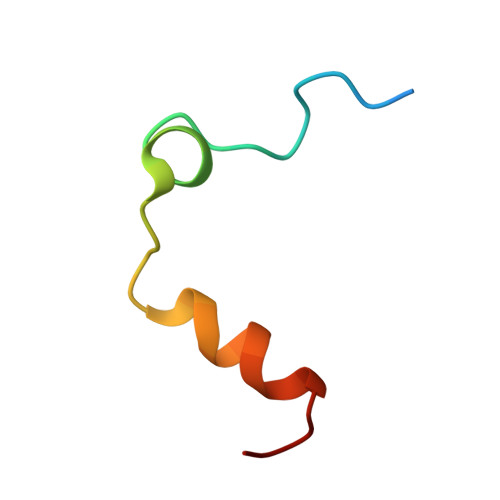
1SFQ, 1SG8, 1SGI, 1SHH - PubMed Abstract:
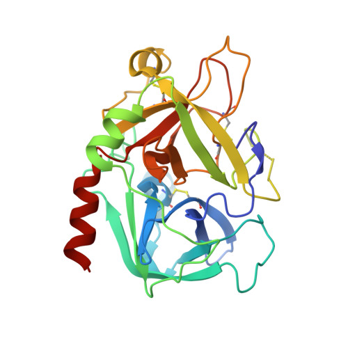
Na(+) binding near the primary specificity pocket of thrombin promotes the procoagulant, prothrombotic, and signaling functions of the enzyme. The effect is mediated allosterically by a communication between the Na(+) site and regions involved in substrate recognition. Using a panel of 78 Ala mutants of thrombin, we have mapped the allosteric core of residues that are energetically linked to Na(+) binding. These residues are Asp-189, Glu-217, Asp-222, and Tyr-225, all in close proximity to the bound Na(+). Among these residues, Asp-189 shares with Asp-221 the important function of transducing Na(+) binding into enhanced catalytic activity. None of the residues of exosite I, exosite II, or the 60-loop plays a significant role in Na(+) binding and allosteric transduction. X-ray crystal structures of the Na(+)-free (slow) and Na(+)-bound (fast) forms of thrombin, free or bound to the active site inhibitor H-d-Phe-Pro-Arg-chloromethyl-ketone, document the conformational changes induced by Na(+) binding. The slow --> fast transition results in formation of the Arg-187:Asp-222 ion pair, optimal orientation of Asp-189 and Ser-195 for substrate binding, and a significant shift of the side chain of Glu-192 linked to a rearrangement of the network of water molecules that connect the bound Na(+) to Ser-195 in the active site. The changes in the water network and the allosteric core explain the thermodynamic signatures linked to Na(+) binding and the mechanism of thrombin activation by Na(+). The role of the water network uncovered in this study establishes a new paradigm for the allosteric regulation of thrombin and other Na(+)-activated enzymes involved in blood coagulation and the immune response.
- Department of Biochemistry and Molecular Biophysics, Washington University School of Medicine, St. Louis, Missouri 63110, USA.
Organizational Affiliation: