1TDI

Crystal Structure of hGSTA3-3 in Complex with Glutathione


Experimental Data Snapshot

  • Method: X-RAY DIFFRACTION
  • Resolution: 2.40 Å
  • R-Value Free: 
    0.252 (Depositor), 0.260 (DCC) 
  • R-Value Work: 
    0.228 (Depositor), 0.240 (DCC) 
  • R-Value Observed: 
    0.228 (Depositor) 

Starting Model: experimental
View more details

wwPDB Validation   3D Report Full Report


Ligand Structure Quality Assessment 

Created with Raphaël 2.3.0Worse 01 BetterLigand structure goodness of fit to experimental dataBest fitted GSHClick on this verticalbar to view details

This is version 1.5 of the entry. See complete history


Literature

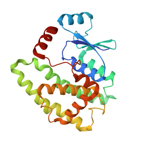
Crystal structure of human glutathione S-transferase A3-3 and mechanistic implications for its high steroid isomerase activity.

Gu, Y.Guo, J.Pal, A.Pan, S.S.Zimniak, P.Singh, S.V.Ji, X.

(2004) Biochemistry 43: 15673-15679

  • DOI: https://doi.org/10.1021/bi048757g
  • Primary Citation of Related Structures:  
    1TDI

  • PubMed Abstract: 

    The crystal structure of human class alpha glutathione (GSH) S-transferase A3-3 (hGSTA3-3) in complex with GSH was determined at 2.4 A. Despite considerable amino acid sequence identity with other human class alpha GSTs (e.g., hGSTA1-1), hGSTA3-3 is unique due to its exceptionally high steroid double bond isomerase activity for the transformation of Delta(5)-androstene-3,17-dione (Delta(5)-AD) to Delta(4)-androstene-3,17-dione. A comparative analysis of the active centers of hGSTA1-1 and hGSTA3-3 reveals that residues in positions 12 and 208 may contribute to their disparate isomerase activity toward Delta(5)-AD. Substitution of these two residues of hGSTA3-3 with the corresponding residues in hGSTA1-1 followed by kinetic characterization of the wild-type and the mutant enzymes supported this prediction. On the basis of our model of the hGSTA3-3.GSH.Delta(5)-AD ternary complex and available biochemical data, we propose that the thiolate group of deprotonated GSH (GS(-)) serves as a base to initiate the reaction by accepting a proton from the steroid and the nonionized hydroxyl group of catalytic residue Y9 (HO-Y9) functions as part of a proton-conducting wire to transfer a proton back to the steroid. Residue R15 may function to stabilize the deprotonated thiolate group of GSH (GS(-)), and a GSH-bound water molecule may donate a hydrogen bond to the 3-keto group of Delta(5)-AD and thus help the thiolate of GS(-) to initiate the proton transfer and the subsequent stabilization of the reaction intermediate.


  • Organizational Affiliation

    Macromolecular Crystallography Laboratory, National Cancer Institute, Frederick, Maryland 21702, USA.


Macromolecules
Find similar proteins by:  (by identity cutoff)  |  3D Structure
Entity ID: 1
MoleculeChains Sequence LengthOrganismDetailsImage
Glutathione S-transferase A3-3
A, B
222Homo sapiensMutation(s): 0 
Gene Names: GSTA3
EC: 2.5.1.18
UniProt & NIH Common Fund Data Resources
Find proteins for Q16772 (Homo sapiens)
Explore Q16772 
Go to UniProtKB:  Q16772
PHAROS:  Q16772
GTEx:  ENSG00000174156 
Entity Groups  
Sequence Clusters30% Identity50% Identity70% Identity90% Identity95% Identity100% Identity
UniProt GroupQ16772
Sequence Annotations
Expand
  • Reference Sequence
Experimental Data & Validation

Experimental Data

  • Method: X-RAY DIFFRACTION
  • Resolution: 2.40 Å
  • R-Value Free:  0.252 (Depositor), 0.260 (DCC) 
  • R-Value Work:  0.228 (Depositor), 0.240 (DCC) 
  • R-Value Observed: 0.228 (Depositor) 
Space Group: P 1 21 1
Unit Cell:
Length ( Å )Angle ( ˚ )
a = 92.985α = 90
b = 95.588β = 90.14
c = 114.039γ = 90
Software Package:
Software NamePurpose
CNSrefinement
DENZOdata reduction
SCALEPACKdata scaling
AMoREphasing

Structure Validation

View Full Validation Report


Ligand Structure Quality Assessment 

Created with Raphaël 2.3.0Worse 01 BetterLigand structure goodness of fit to experimental dataBest fitted GSHClick on this verticalbar to view details

Entry History 

Deposition Data

Revision History  (Full details and data files)

  • Version 1.0: 2005-01-18
    Type: Initial release
  • Version 1.1: 2008-04-30
    Changes: Version format compliance
  • Version 1.2: 2011-07-13
    Changes: Version format compliance
  • Version 1.3: 2014-09-17
    Changes: Other
  • Version 1.4: 2023-08-23
    Changes: Data collection, Database references, Derived calculations, Refinement description
  • Version 1.5: 2023-08-30
    Changes: Database references, Structure summary