1UA7 | pdb_00001ua7

Crystal Structure Analysis of Alpha-Amylase from Bacillus Subtilis complexed with Acarbose


Experimental Data Snapshot

  • Method: X-RAY DIFFRACTION
  • Resolution: 2.21 Å
  • R-Value Free: 
    0.265 (Depositor), 0.258 (DCC) 
  • R-Value Work: 
    0.208 (Depositor), 0.201 (DCC) 
  • R-Value Observed: 
    0.208 (Depositor) 

wwPDB Validation 3D Report Full Report

Validation slider image for 1UA7

Ligand Structure Quality Assessment 


This is version 3.2 of the entry. See complete history

Literature

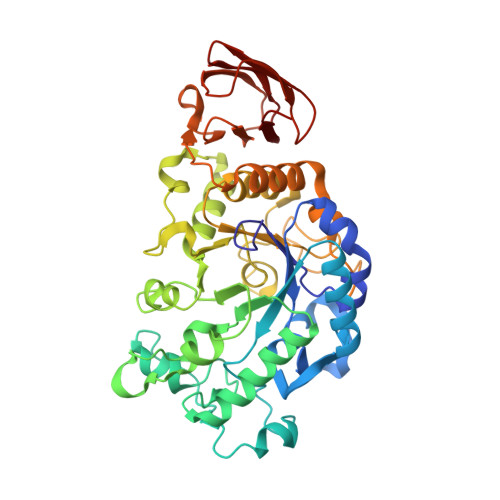
Crystal structure of Bacillus subtilis alpha-amylase in complex with acarbose

Kagawa, M.Fujimoto, Z.Momma, M.Takase, K.Mizuno, H.

(2003) J Bacteriol 185: 6981-6984

  • DOI: https://doi.org/10.1128/JB.185.23.6981-6984.2003
  • Primary Citation Related Structures: 
    1UA7

  • PubMed Abstract: 

    The crystal structure of Bacillus subtilis alpha-amylase, in complex with the pseudotetrasaccharide inhibitor acarbose, revealed an hexasaccharide in the active site as a result of transglycosylation. After comparison with the known structure of the catalytic-site mutant complexed with the native substrate maltopentaose, it is suggested that the present structure represents a mimic intermediate in the initial stage of the catalytic process.


  • Organizational Affiliation
    • Department of Biochemistry, National Institute of Agrobiological Sciences, Tsukuba, Ibaraki 305-8602, Japan.

Macromolecule Content 

  • Total Structure Weight: 47.9 kDa 
  • Atom Count: 3,808 
  • Modeled Residue Count: 422 
  • Deposited Residue Count: 422 
  • Unique protein chains: 1

Macromolecules

Find similar proteins by:|  3D Structure
Entity ID: 1
MoleculeChains  Sequence LengthOrganismDetailsImage
Alpha-amylase422Bacillus subtilisMutation(s): 1 
EC: 3.2.1.1
UniProt
Find proteins for P00691 (Bacillus subtilis (strain 168))
Explore P00691 
Go to UniProtKB:  P00691
Entity Groups
Sequence Clusters30% Identity50% Identity70% Identity90% Identity95% Identity100% Identity
UniProt GroupP00691
Sequence Annotations
Expand
Reference Sequence

Oligosaccharides

Help  
Entity ID: 2
MoleculeChains Length2D Diagram GlycosylationD Interactions
4,6-dideoxy-alpha-D-xylo-hexopyranose-(1-4)-alpha-D-glucopyranose
B
2N/A
Glycosylation Resources
GlyTouCan: G59234JH
GlyCosmos: G59234JH
Entity ID: 3
MoleculeChains Length2D Diagram GlycosylationD Interactions
alpha-D-quinovopyranose-(1-4)-beta-D-glucopyranose
C
2N/A
Glycosylation Resources
GlyTouCan: G40192QI
GlyCosmos: G40192QI
GlyGen: G40192QI

Experimental Data & Validation

Experimental Data

  • Method: X-RAY DIFFRACTION
  • Resolution: 2.21 Å
  • R-Value Free:  0.265 (Depositor), 0.258 (DCC) 
  • R-Value Work:  0.208 (Depositor), 0.201 (DCC) 
  • R-Value Observed: 0.208 (Depositor) 
Space Group: P 21 21 21
Unit Cell:
Length ( Å )Angle ( ˚ )
a = 70.32α = 90
b = 74.204β = 90
c = 115.724γ = 90
Software Package:
Software NamePurpose
CNSrefinement

Structure Validation

View Full Validation Report


Ligand Structure Quality Assessment 


Entry History 

Deposition Data

Revision History  (Full details and data files)

  • Version 1.0: 2004-05-18
    Type: Initial release
  • Version 1.1: 2008-04-27
    Changes: Version format compliance
  • Version 1.2: 2011-07-13
    Changes: Version format compliance
  • Version 1.3: 2017-10-04
    Changes: Refinement description
  • Version 2.0: 2017-10-25
    Changes: Atomic model, Derived calculations, Structure summary
  • Version 3.0: 2020-07-29
    Type: Remediation
    Reason: Carbohydrate remediation
    Changes: Atomic model, Data collection, Derived calculations, Structure summary
  • Version 3.1: 2021-11-10
    Changes: Database references, Structure summary
  • Version 3.2: 2023-12-27
    Changes: Data collection