1UMA

ALPHA-THROMBIN (HIRUGEN) COMPLEXED WITH NA-(N,N-DIMETHYLCARBAMOYL)-ALPHA-AZALYSINE


Experimental Data Snapshot

  • Method: X-RAY DIFFRACTION
  • Resolution: 2.00 Å
  • R-Value Work: 
    0.177 (DCC) 

wwPDB Validation   3D Report Full Report


Ligand Structure Quality Assessment 

Created with Raphaël 2.3.0Worse 01 BetterLigand structure goodness of fit to experimental dataBest fitted NAGClick on this verticalbar to view detailsBest fitted IN2Click on this verticalbar to view details

This is version 1.5 of the entry. See complete history


Literature

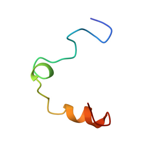
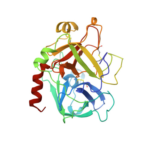
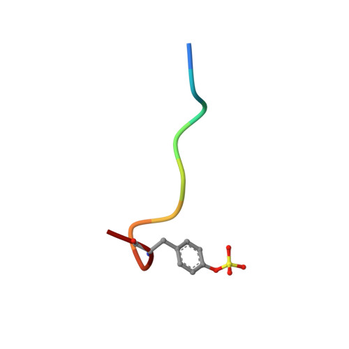
Human alpha-thrombin inhibition by the active site titrant N alpha-(N,N-dimethylcarbamoyl)-alpha-azalysine p-nitrophenyl ester: a comparative kinetic and X-ray crystallographic study.

Nardini, M.Pesce, A.Rizzi, M.Casale, E.Ferraccioli, R.Balliano, G.Milla, P.Ascenzi, P.Bolognesi, M.

(1996) J Mol Biol 258: 851-859

  • DOI: https://doi.org/10.1006/jmbi.1996.0292
  • Primary Citation of Related Structures:  
    1UMA

  • PubMed Abstract: 

    Kinetics for the hydrolysis of the chromogenic active site titrant N alpha-(N,N-dimethylcarbamoyl)-alpha-azalysine p-nitrophenyl ester (Dmc-azaLys-ONp) catalyzed by bovine beta-trypsin, bovine alpha-thrombin, human alpha-thrombin, human Lys77-plasmin, human urinary kallikrein, the M(r) 33,000 and M(r) 54,000 species of human urokinase, as well as by porcine pancreatic beta-kallikrein-A and B have been obtained between pH 6.0 and 8.0, at 21.0 degrees C. Moreover, the three dimensional structure of the human alpha-thrombin-(hirugen).Dmc-azaLys acyl.enzyme complex has been analyzed and refined by X-ray crystallography at 2.0 A resolution (R-factor = 0.168). As observed for bovine beta-trypsin, the acylating inhibitor molecule is covalently bound to the Ser195 catalytic residue, filling the human alpha-thrombin S1 primary specificity subsite with its lysyl side-group. However, the carbonyl group of the scissile human alpha-thrombin.Dmc-azaLys acyl bond does not occupy properly the oxyanion binding hole. At variance from the bovine beta-trypsin.Dmc-azaLys acyl.enzyme structure, a second, not covalently bound, inhibitor molecule, partly shielded by the 60-insertion loop of human alpha-thrombin, is contacting the enzyme "aryl-binding site".


  • Organizational Affiliation

    Centro Biotecnologie Avanzate IST, Università di Genova, Italy.


Macromolecules
Find similar proteins by:  (by identity cutoff)  |  3D Structure
Entity ID: 1
MoleculeChains Sequence LengthOrganismDetailsImage
ALPHA-THROMBINA [auth L]36Homo sapiensMutation(s): 0 
EC: 3.4.21.5
UniProt & NIH Common Fund Data Resources
Find proteins for P00734 (Homo sapiens)
Explore P00734 
Go to UniProtKB:  P00734
PHAROS:  P00734
GTEx:  ENSG00000180210 
Entity Groups  
Sequence Clusters30% Identity50% Identity70% Identity90% Identity95% Identity100% Identity
UniProt GroupP00734
Sequence Annotations
Expand
  • Reference Sequence
Find similar proteins by:  (by identity cutoff)  |  3D Structure
Entity ID: 2
MoleculeChains Sequence LengthOrganismDetailsImage
ALPHA-THROMBINB [auth H]259Homo sapiensMutation(s): 0 
EC: 3.4.21.5
UniProt & NIH Common Fund Data Resources
Find proteins for P00734 (Homo sapiens)
Explore P00734 
Go to UniProtKB:  P00734
PHAROS:  P00734
GTEx:  ENSG00000180210 
Entity Groups  
Sequence Clusters30% Identity50% Identity70% Identity90% Identity95% Identity100% Identity
UniProt GroupP00734
Glycosylation
Glycosylation Sites: 1Go to GlyGen: P00734-1
Sequence Annotations
Expand
  • Reference Sequence

Find similar proteins by:  Sequence   |   3D Structure  

Entity ID: 3
MoleculeChains Sequence LengthOrganismDetailsImage
HIRUDIN IC [auth I]10Hirudo medicinalisMutation(s): 0 
UniProt
Find proteins for P01050 (Hirudo medicinalis)
Explore P01050 
Go to UniProtKB:  P01050
Entity Groups  
Sequence Clusters30% Identity50% Identity70% Identity90% Identity95% Identity100% Identity
UniProt GroupP01050
Sequence Annotations
Expand
  • Reference Sequence
Small Molecules
Modified Residues  1 Unique
IDChains TypeFormula2D DiagramParent
TYS
Query on TYS
C [auth I]L-PEPTIDE LINKINGC9 H11 N O6 STYR
Experimental Data & Validation

Experimental Data

  • Method: X-RAY DIFFRACTION
  • Resolution: 2.00 Å
  • R-Value Work:  0.177 (DCC) 
Space Group: C 1 2 1
Unit Cell:
Length ( Å )Angle ( ˚ )
a = 71.15α = 90
b = 71.9β = 100.49
c = 72.3γ = 90
Software Package:
Software NamePurpose
MOSFLMdata reduction
TNTrefinement

Structure Validation

View Full Validation Report


Ligand Structure Quality Assessment 

Created with Raphaël 2.3.0Worse 01 BetterLigand structure goodness of fit to experimental dataBest fitted NAGClick on this verticalbar to view detailsBest fitted IN2Click on this verticalbar to view details

Entry History 

Deposition Data

Revision History  (Full details and data files)

  • Version 1.0: 1996-11-08
    Type: Initial release
  • Version 1.1: 2008-03-03
    Changes: Version format compliance
  • Version 1.2: 2011-07-13
    Changes: Atomic model, Database references, Derived calculations, Non-polymer description, Structure summary, Version format compliance
  • Version 1.3: 2011-11-16
    Changes: Atomic model
  • Version 1.4: 2020-07-29
    Type: Remediation
    Reason: Carbohydrate remediation
    Changes: Advisory, Data collection, Derived calculations, Structure summary
  • Version 1.5: 2024-10-09
    Changes: Advisory, Data collection, Database references, Structure summary