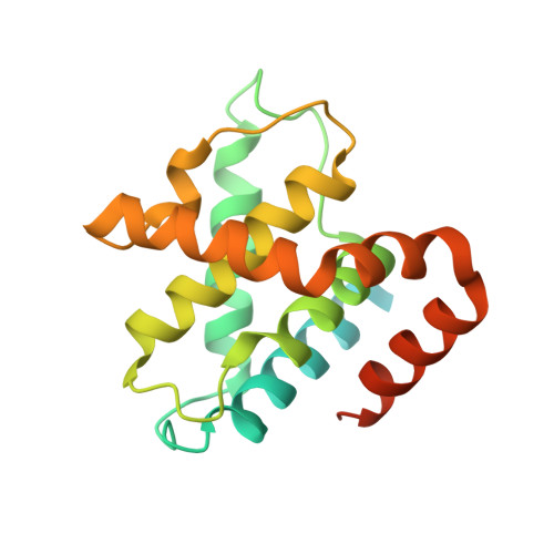
1UNH

Structural mechanism for the inhibition of CDK5-p25 by roscovitine, aloisine and indirubin.


Experimental Data Snapshot

  • Method: X-RAY DIFFRACTION
  • Resolution: 2.35 Å
  • R-Value Free: 0.230 
  • R-Value Work: 0.229 
  • R-Value Observed: 0.229 

Starting Model: experimental
View more details

wwPDB Validation   3D Report Full Report


Ligand Structure Quality Assessment 


This is version 1.2 of the entry. See complete history


Literature

Mechanism of Cdk5/P25 Binding by Cdk Inhibitors

Mapelli, M.Massimilinao, L.Crovace, C.Seeliger, M.A.Tsai, L.-H.Meijer, L.Musacchio, A.

(2005) J Med Chem 48: 671

  • DOI: https://doi.org/10.1021/jm049323m
  • Primary Citation of Related Structures:  
    1UNG, 1UNH, 1UNL

  • PubMed Abstract: 

    The cyclin-dependent kinases (CDK) CDK1, CDK2, CDK4, and CDK6 are serine/threonine protein kinases targeted in cancer therapy due to their role in cell cycle progression. The postmitotic CDK5 is involved in biological pathways important for neuronal migration and differentiation. CDK5 represents an attractive pharmacological target as its deregulation is implicated in various neurodegenerative diseases such as Alzheimer's, Parkinson's, and Niemann-Pick type C diseases, ischemia, and amyotrophic lateral sclerosis. We have generated an improved crystal form of CDK5 in complex with p25, a segment of the p35 neuronal activator. The crystals were used to solve the structure of CDK5/p25 with (R)-roscovitine and aloisine at a resolution of 2.2 and 2.3 A, respectively. The structure of CDK5/p25/roscovitine provides a rationale for the preference of CDK5 for the R over the S stereoisomer. Furthermore, roscovitine stabilized an unusual collapsed conformation of the glycine-rich loop, an important site of CDK regulation, and we report an investigation of the effects of glycine-rich loop phosphorylation on roscovitine binding. The CDK5/p25 crystals represent a valuable new tool for the identification and optimization of selective CDK inhibitors.


  • Organizational Affiliation

    Structural Biology Unit, Department of Experimental Oncology, European Institute of Oncology, Via Ripamonti 435, 20141 Milan, Italy.


Macromolecules
Find similar proteins by:  (by identity cutoff)  |  3D Structure
Entity ID: 1
MoleculeChains Sequence LengthOrganismDetailsImage
CYCLIN-DEPENDENT KINASE 5
A, B
292Homo sapiensMutation(s): 1 
EC: 2.7.11.22 (PDB Primary Data), 2.7.11.1 (UniProt)
UniProt & NIH Common Fund Data Resources
Find proteins for Q00535 (Homo sapiens)
Explore Q00535 
Go to UniProtKB:  Q00535
PHAROS:  Q00535
GTEx:  ENSG00000164885 
Entity Groups  
Sequence Clusters30% Identity50% Identity70% Identity90% Identity95% Identity100% Identity
UniProt GroupQ00535
Sequence Annotations
Expand
  • Reference Sequence
Find similar proteins by:  (by identity cutoff)  |  3D Structure
Entity ID: 2
MoleculeChains Sequence LengthOrganismDetailsImage
CYCLIN-DEPENDENT KINASE 5 ACTIVATOR 1C [auth D],
D [auth E]
208Homo sapiensMutation(s): 0 
UniProt & NIH Common Fund Data Resources
Find proteins for Q15078 (Homo sapiens)
Explore Q15078 
Go to UniProtKB:  Q15078
PHAROS:  Q15078
GTEx:  ENSG00000176749 
Entity Groups  
Sequence Clusters30% Identity50% Identity70% Identity90% Identity95% Identity100% Identity
UniProt GroupQ15078
Sequence Annotations
Expand
  • Reference Sequence
Small Molecules
Binding Affinity Annotations 
IDSourceBinding Affinity
IXM BindingDB:  1UNH IC50: 100 (nM) from 1 assay(s)
PDBBind:  1UNH IC50: 100 (nM) from 1 assay(s)
Experimental Data & Validation

Experimental Data

  • Method: X-RAY DIFFRACTION
  • Resolution: 2.35 Å
  • R-Value Free: 0.230 
  • R-Value Work: 0.229 
  • R-Value Observed: 0.229 
  • Space Group: C 1 2 1
Unit Cell:
Length ( Å )Angle ( ˚ )
a = 149.541α = 90
b = 90.124β = 93.29
c = 83.158γ = 90
Software Package:
Software NamePurpose
REFMACrefinement
DENZOdata reduction
SCALEPACKdata scaling
AMoREphasing

Structure Validation

View Full Validation Report


Ligand Structure Quality Assessment 


Entry History 

Deposition Data

Revision History  (Full details and data files)

  • Version 1.0: 2004-11-10
    Type: Initial release
  • Version 1.1: 2011-07-13
    Changes: Advisory, Version format compliance
  • Version 1.2: 2023-12-13
    Changes: Data collection, Database references, Other, Refinement description