1UZU

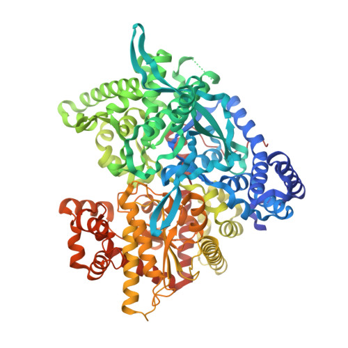
Glycogen Phosphorylase b in complex with indirubin-5'-sulphonate


Experimental Data Snapshot

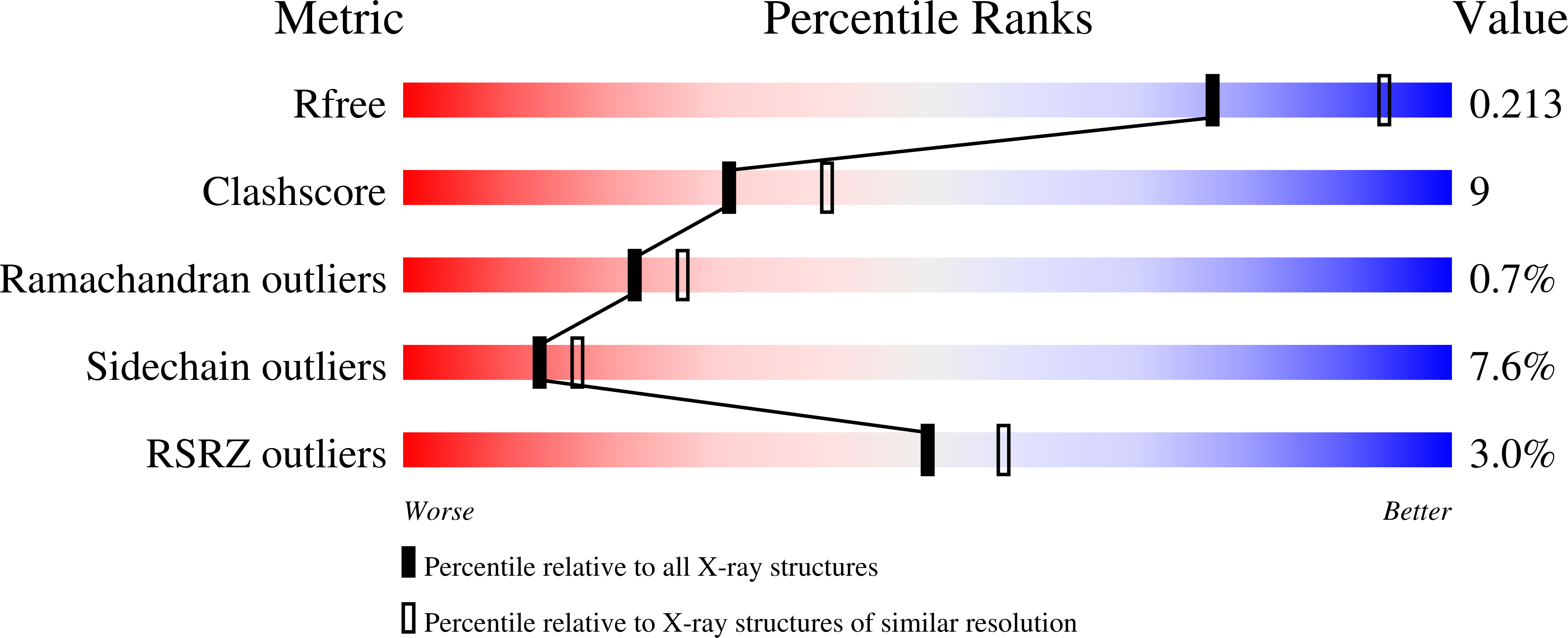
  • Method: X-RAY DIFFRACTION
  • Resolution: 2.30 Å
  • R-Value Free: 0.214 
  • R-Value Work: 0.182 
  • R-Value Observed: 0.182 

Starting Model: experimental
View more details

wwPDB Validation   3D Report Full Report


Ligand Structure Quality Assessment 


This is version 1.4 of the entry. See complete history


Literature

Binding of the potential antitumour agent indirubin-5-sulphonate at the inhibitor site of rabbit muscle glycogen phosphorylase b. Comparison with ligand binding to pCDK2-cyclin A complex.

Kosmopoulou, M.N.Leonidas, D.D.Chrysina, E.D.Bischler, N.Eisenbrand, G.Sakarellos, C.E.Pauptit, R.Oikonomakos, N.G.

(2004) Eur J Biochem 271: 2280-2290

  • DOI: https://doi.org/10.1111/j.1432-1033.2004.04173.x
  • Primary Citation of Related Structures:  
    1UZU

  • PubMed Abstract: 

    The binding of indirubin-5-sulphonate (E226), a potential anti-tumour agent and a potent inhibitor (IC(50) = 35 nm) of cyclin-dependent kinase 2 (CDK2) and glycogen phosphorylase (GP) has been studied by kinetic and crystallographic methods. Kinetic analysis revealed that E226 is a moderate inhibitor of GPb (K(i) = 13.8 +/- 0.2 micro m) and GPa (K(i) = 57.8 +/- 7.1 micro m) and acts synergistically with glucose. To explore the molecular basis of E226 binding we have determined the crystal structure of the GPb/E226 complex at 2.3 A resolution. Structure analysis shows clearly that E226 binds at the purine inhibitor site, where caffeine and flavopiridol also bind [Oikonomakos, N.G., Schnier, J.B., Zographos, S.E., Skamnaki, V.T., Tsitsanou, K.E. & Johnson, L.N. (2000) J. Biol. Chem.275, 34566-34573], by intercalating between the two aromatic rings of Phe285 and Tyr613. The mode of binding of E226 to GPb is similar, but not identical, to that of caffeine and flavopiridol. Comparative structural analyses of the GPb-E226, GPb-caffeine and GPb-flavopiridol complex structures reveal the structural basis of the differences in the potencies of the three inhibitors and indicate binding residues in the inhibitor site that can be exploited to obtain more potent inhibitors. Structural comparison of the GPb-E226 complex structure with the active pCDK2-cyclin A-E226 complex structure clearly shows the different binding modes of the ligand to GPb and CDK2; the more extensive interactions of E226 with the active site of CDK2 may explain its higher affinity towards the latter enzyme.


  • Organizational Affiliation

    Institute of Organic and Pharmaceutical Chemistry, The National Hellenic Research Foundation, 48 Vas. Constantinou Avenue, 11635 Athens, Greece.


Macromolecules
Find similar proteins by:  (by identity cutoff)  |  3D Structure
Entity ID: 1
MoleculeChains Sequence LengthOrganismDetailsImage
GLYCOGEN PHOSPHORYLASE, MUSCLE FORM842Oryctolagus cuniculusMutation(s): 0 
EC: 2.4.1.1
UniProt
Find proteins for P00489 (Oryctolagus cuniculus)
Explore P00489 
Go to UniProtKB:  P00489
Entity Groups  
Sequence Clusters30% Identity50% Identity70% Identity90% Identity95% Identity100% Identity
UniProt GroupP00489
Sequence Annotations
Expand
  • Reference Sequence
Small Molecules
Ligands 2 Unique
IDChains Name / Formula / InChI Key2D Diagram3D Interactions
INR
Query on INR

Download Ideal Coordinates CCD File 
C [auth A]2',3-DIOXO-1,1',2',3-TETRAHYDRO-2,3'-BIINDOLE-5'-SULFONIC ACID
C16 H10 N2 O5 S
IHBOEHLUIBMBMY-YPKPFQOOSA-N
PLP
Query on PLP

Download Ideal Coordinates CCD File 
B [auth A]PYRIDOXAL-5'-PHOSPHATE
C8 H10 N O6 P
NGVDGCNFYWLIFO-UHFFFAOYSA-N
Experimental Data & Validation

Experimental Data

  • Method: X-RAY DIFFRACTION
  • Resolution: 2.30 Å
  • R-Value Free: 0.214 
  • R-Value Work: 0.182 
  • R-Value Observed: 0.182 
  • Space Group: P 43 21 2
Unit Cell:
Length ( Å )Angle ( ˚ )
a = 128.652α = 90
b = 128.652β = 90
c = 116.389γ = 90
Software Package:
Software NamePurpose
CNSrefinement
DENZOdata reduction
SCALEPACKdata scaling
CNSphasing

Structure Validation

View Full Validation Report


Ligand Structure Quality Assessment 


Entry History 

Deposition Data

Revision History  (Full details and data files)

  • Version 1.0: 2004-05-27
    Type: Initial release
  • Version 1.1: 2011-05-07
    Changes: Version format compliance
  • Version 1.2: 2011-07-13
    Changes: Version format compliance
  • Version 1.3: 2019-02-06
    Changes: Data collection, Database references, Experimental preparation
  • Version 1.4: 2023-12-13
    Changes: Data collection, Database references, Derived calculations, Other, Refinement description