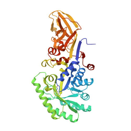
1VFS

Crystal structure of D-cycloserine-bound form of alanine racemase from D-cycloserine-producing Streptomyces lavendulae


Experimental Data Snapshot

  • Method: X-RAY DIFFRACTION
  • Resolution: 1.90 Å
  • R-Value Free: 0.265 
  • R-Value Work: 0.209 
  • R-Value Observed: 0.209 

Starting Model: experimental
View more details

wwPDB Validation   3D Report Full Report


This is version 1.4 of the entry. See complete history


Literature

Structural evidence that alanine racemase from a D-cycloserine-producing microorganism exhibits resistance to its own product.

Noda, M.Matoba, Y.Kumagai, T.Sugiyama, M.

(2004) J Biol Chem 279: 46153-46161

  • DOI: https://doi.org/10.1074/jbc.M404605200
  • Primary Citation of Related Structures:  
    1VFH, 1VFS, 1VFT

  • PubMed Abstract: 

    Alanine racemase (ALR), an enzyme that catalyzes the interconversion of Ala enantiomers, is essential for the synthesis of the bacterial cell wall. We have shown that it is harder to inhibit the catalytic activity of ALR from D-cycloserine (DCS)-producing Streptomyces lavendulae than that from Escherichia coli by DCS. To obtain structural evidence for the fact that Streptomyces ALR displays resistance to DCS, we determined the precise nature of the x-ray crystal structures of the cycloserine-free and cycloserine enantiomer-bound forms of Streptomyces ALR at high resolutions. Streptomyces ALR takes a dimer structure, which is formed by interactions between the N-terminal domain of one monomer with the C-terminal domain of its partner. Each of the two active sites of ALR, which is generated as a result of the formation of the dimer structure, is composed of pyridoxal 5'-phosphate (PLP), the PLP-binding residue Lys(38), and the amino acids in the immediate environment of the pyridoxal cofactor. The current model suggests that each active site of Streptomyces ALR maintains a larger space and takes a more rigid conformation than that of Bacillus stearothermophilus ALR determined previously. Furthermore, we show that Streptomyces ALR results in a slow conversion to a final form of a pyridoxal derivative arising from either isomer of cycloserine, which inhibits the catalytic activity noncompetitively. In fact, the slow conversion is confirmed by the fact that each enzyme bound cycloserine derivative, which is bound to PLP, takes an asymmetric structure.


  • Organizational Affiliation

    Department of Molecular Microbiology and Biotechnology, Graduate School of Biomedical Sciences, Hiroshima University, Kasumi 1-2-3, Minami-Ku, Hiroshima 734-8551, Japan.


Macromolecules
Find similar proteins by:  (by identity cutoff)  |  3D Structure
Entity ID: 1
MoleculeChains Sequence LengthOrganismDetailsImage
alanine racemase
A, B
386Streptomyces lavendulaeMutation(s): 0 
Gene Names: alr
EC: 5.1.1.1
UniProt
Find proteins for Q65YW7 (Streptomyces lavendulae subsp. lavendulae)
Explore Q65YW7 
Go to UniProtKB:  Q65YW7
Entity Groups  
Sequence Clusters30% Identity50% Identity70% Identity90% Identity95% Identity100% Identity
UniProt GroupQ65YW7
Sequence Annotations
Expand
  • Reference Sequence
Small Molecules
Ligands 2 Unique
IDChains Name / Formula / InChI Key2D Diagram3D Interactions
DCS
Query on DCS

Download Ideal Coordinates CCD File 
D [auth A],
E [auth B]
D-[3-HYDROXY-2-METHYL-5-PHOSPHONOOXYMETHYL-PYRIDIN-4-YLMETHYL]-N,O-CYCLOSERYLAMIDE
C11 H16 N3 O7 P
NNRZSZJOQKAGTO-SECBINFHSA-N
CL
Query on CL

Download Ideal Coordinates CCD File 
C [auth A]CHLORIDE ION
Cl
VEXZGXHMUGYJMC-UHFFFAOYSA-M
Modified Residues  1 Unique
IDChains TypeFormula2D DiagramParent
KCX
Query on KCX
A, B
L-PEPTIDE LINKINGC7 H14 N2 O4LYS
Experimental Data & Validation

Experimental Data

  • Method: X-RAY DIFFRACTION
  • Resolution: 1.90 Å
  • R-Value Free: 0.265 
  • R-Value Work: 0.209 
  • R-Value Observed: 0.209 
  • Space Group: P 1 21 1
Unit Cell:
Length ( Å )Angle ( ˚ )
a = 82.96α = 90
b = 63.55β = 118.61
c = 84.62γ = 90
Software Package:
Software NamePurpose
X-PLORrefinement
MOSFLMdata reduction
CCP4data scaling
AMoREphasing

Structure Validation

View Full Validation Report


Entry History 

Deposition Data

Revision History  (Full details and data files)

  • Version 1.0: 2004-09-14
    Type: Initial release
  • Version 1.1: 2008-04-27
    Changes: Version format compliance
  • Version 1.2: 2011-07-13
    Changes: Version format compliance
  • Version 1.3: 2023-10-25
    Changes: Data collection, Database references, Derived calculations, Refinement description
  • Version 1.4: 2023-11-15
    Changes: Data collection