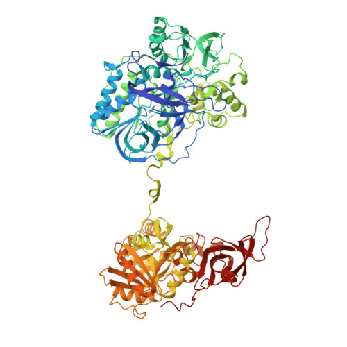
1VRQ

Crystal Structure of Heterotetrameric Sarcosine Oxidase from Corynebacterium sp. U-96 in complex with Folinic Acid


Experimental Data Snapshot

  • Method: X-RAY DIFFRACTION
  • Resolution: 2.20 Å
  • R-Value Free: 0.224 
  • R-Value Work: 0.192 
  • R-Value Observed: 0.192 

wwPDB Validation   3D Report Full Report


Ligand Structure Quality Assessment 


This is version 1.3 of the entry. See complete history


Literature

Crystal structure of heterotetrameric sarcosine oxidase from Corynebacterium sp. U-96

Ida, K.Moriguchi, T.Suzuki, H.

(2005) Biochem Biophys Res Commun 333: 359-366

  • DOI: https://doi.org/10.1016/j.bbrc.2005.05.116
  • Primary Citation of Related Structures:  
    1VRQ, 1X31

  • PubMed Abstract: 

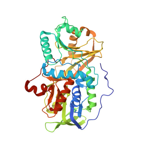
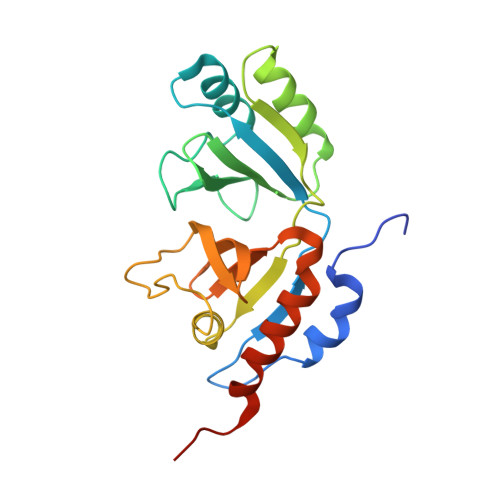
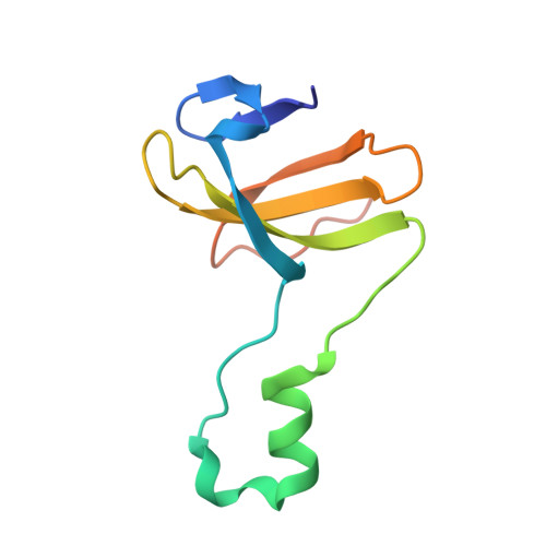
    Sarcosine oxidase from Corynebacterium sp. U-96 is a heterotetrameric enzyme. Here we report the crystal structures of the enzyme in complex with dimethylglycine and folinic acid. The alpha subunit is composed of two domains, contains NAD(+), and binds folinic acid. The beta subunit contains dimethylglycine, FAD, and FMN, and these flavins are approximately 10A apart. The gamma subunit is in contact with two domains of alpha subunit and has possibly a folate-binding structure. The delta subunit contains a single atom of zinc and has a Cys(3)His zinc finger structure. Based on the structures determined and on the previous works, the structure-function relationship on the heterotetrameric sarcosine oxidase is discussed.


  • Organizational Affiliation

    Department of Biosciences, School of Science, Kitasato University, 1-15-1 Kitasato, Sagamihara, Kanagawa 228-8555, Japan. idakoh@sci.kitasato-u.ac.jp


Macromolecules
Find similar proteins by:  (by identity cutoff)  |  3D Structure
Entity ID: 1
MoleculeChains Sequence LengthOrganismDetailsImage
Sarcosine oxidase alpha subunit964Corynebacterium sp. U-96Mutation(s): 0 
EC: 1.5.3.1 (PDB Primary Data), 1.5.3.24 (UniProt)
UniProt
Find proteins for Q50LF0 (Corynebacterium sp. (strain U-96))
Explore Q50LF0 
Go to UniProtKB:  Q50LF0
Entity Groups  
Sequence Clusters30% Identity50% Identity70% Identity90% Identity95% Identity100% Identity
UniProt GroupQ50LF0
Sequence Annotations
Expand
  • Reference Sequence
Find similar proteins by:  (by identity cutoff)  |  3D Structure
Entity ID: 2
MoleculeChains Sequence LengthOrganismDetailsImage
Sarcosine oxidase beta subunit404Corynebacterium sp. U-96Mutation(s): 0 
EC: 1.5.3.1 (PDB Primary Data), 1.5.3.24 (UniProt)
UniProt
Find proteins for Q50LF2 (Corynebacterium sp. (strain U-96))
Explore Q50LF2 
Go to UniProtKB:  Q50LF2
Entity Groups  
Sequence Clusters30% Identity50% Identity70% Identity90% Identity95% Identity100% Identity
UniProt GroupQ50LF2
Sequence Annotations
Expand
  • Reference Sequence
Find similar proteins by:  (by identity cutoff)  |  3D Structure
Entity ID: 3
MoleculeChains Sequence LengthOrganismDetailsImage
Sarcosine oxidase gamma subunit206Corynebacterium sp. U-96Mutation(s): 0 
EC: 1.5.3.1 (PDB Primary Data), 1.5.3.24 (UniProt)
UniProt
Find proteins for Q50LE9 (Corynebacterium sp. (strain U-96))
Explore Q50LE9 
Go to UniProtKB:  Q50LE9
Entity Groups  
Sequence Clusters30% Identity50% Identity70% Identity90% Identity95% Identity100% Identity
UniProt GroupQ50LE9
Sequence Annotations
Expand
  • Reference Sequence
Find similar proteins by:  (by identity cutoff)  |  3D Structure
Entity ID: 4
MoleculeChains Sequence LengthOrganismDetailsImage
Sarcosine oxidase delta subunit99Corynebacterium sp. U-96Mutation(s): 0 
EC: 1.5.3.1 (PDB Primary Data), 1.5.3.24 (UniProt)
UniProt
Find proteins for Q50LF1 (Corynebacterium sp. (strain U-96))
Explore Q50LF1 
Go to UniProtKB:  Q50LF1
Entity Groups  
Sequence Clusters30% Identity50% Identity70% Identity90% Identity95% Identity100% Identity
UniProt GroupQ50LF1
Sequence Annotations
Expand
  • Reference Sequence
Small Molecules
Ligands 7 Unique
IDChains Name / Formula / InChI Key2D Diagram3D Interactions
FAD
Query on FAD

Download Ideal Coordinates CCD File 
Q [auth B]FLAVIN-ADENINE DINUCLEOTIDE
C27 H33 N9 O15 P2
VWWQXMAJTJZDQX-UYBVJOGSSA-N
NAD
Query on NAD

Download Ideal Coordinates CCD File 
M [auth A]NICOTINAMIDE-ADENINE-DINUCLEOTIDE
C21 H27 N7 O14 P2
BAWFJGJZGIEFAR-NNYOXOHSSA-N
FON
Query on FON

Download Ideal Coordinates CCD File 
N [auth A]N-{[4-({[(6R)-2-amino-5-formyl-4-oxo-1,4,5,6,7,8-hexahydropteridin-6-yl]methyl}amino)phenyl]carbonyl}-L-glutamic acid
C20 H23 N7 O7
VVIAGPKUTFNRDU-OLZOCXBDSA-N
FMN
Query on FMN

Download Ideal Coordinates CCD File 
R [auth B]FLAVIN MONONUCLEOTIDE
C17 H21 N4 O9 P
FVTCRASFADXXNN-SCRDCRAPSA-N
DMG
Query on DMG

Download Ideal Coordinates CCD File 
S [auth B]N,N-DIMETHYLGLYCINE
C4 H9 N O2
FFDGPVCHZBVARC-UHFFFAOYSA-N
SO4
Query on SO4

Download Ideal Coordinates CCD File 
E [auth A]
F [auth A]
G [auth A]
H [auth A]
I [auth A]
E [auth A],
F [auth A],
G [auth A],
H [auth A],
I [auth A],
J [auth A],
K [auth A],
L [auth A],
O [auth B],
P [auth B],
T [auth C],
V [auth D],
W [auth D]
SULFATE ION
O4 S
QAOWNCQODCNURD-UHFFFAOYSA-L
ZN
Query on ZN

Download Ideal Coordinates CCD File 
U [auth D]ZINC ION
Zn
PTFCDOFLOPIGGS-UHFFFAOYSA-N
Experimental Data & Validation

Experimental Data

  • Method: X-RAY DIFFRACTION
  • Resolution: 2.20 Å
  • R-Value Free: 0.224 
  • R-Value Work: 0.192 
  • R-Value Observed: 0.192 
  • Space Group: P 65 2 2
Unit Cell:
Length ( Å )Angle ( ˚ )
a = 199.4017α = 90
b = 199.4017β = 90
c = 197.3089γ = 120
Software Package:
Software NamePurpose
MOSFLMdata reduction
SCALAdata scaling
CNSrefinement
CCP4data scaling
CNSphasing

Structure Validation

View Full Validation Report


Ligand Structure Quality Assessment 


Entry History 

Deposition Data

Revision History  (Full details and data files)

  • Version 1.0: 2005-07-05
    Type: Initial release
  • Version 1.1: 2008-04-27
    Changes: Version format compliance
  • Version 1.2: 2011-07-13
    Changes: Source and taxonomy, Version format compliance
  • Version 1.3: 2023-12-27
    Changes: Data collection, Database references, Derived calculations