1W4O | pdb_00001w4o

Binding of Nonnatural 3'-Nucleotides to Ribonuclease A


Experimental Data Snapshot

  • Method: X-RAY DIFFRACTION
  • Resolution: 1.60 Å
  • R-Value Free: 
    0.248 (Depositor) 
  • R-Value Work: 
    0.232 (Depositor), 0.235 (DCC) 
  • R-Value Observed: 
    0.232 (Depositor) 

Starting Model: experimental
View more details

wwPDB Validation 3D Report Full Report

Validation slider image for 1W4O

Ligand Structure Quality Assessment 


This is version 1.4 of the entry. See complete history

Literature

Binding of Non-Natural 3'-Nucleotides to Ribonuclease A

Jenkins, C.L.Thiyagarajan, N.Sweeney, R.Y.Guy, M.P.Kelemen, B.R.Acharya, K.R.Raines, R.T.

(2005) FEBS J 272: 744

  • DOI: https://doi.org/10.1111/j.1742-4658.2004.04511.x
  • Primary Citation Related Structures: 
    1W4O, 1W4P, 1W4Q

  • PubMed Abstract: 

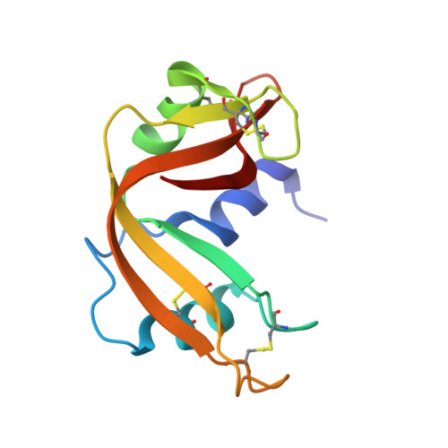
    2'-Fluoro-2'-deoxyuridine 3'-phosphate (dU(F)MP) and arabinouridine 3'-phosphate (araUMP) have non-natural furanose rings. dU(F)MP and araUMP were prepared by chemical synthesis and found to have three- to sevenfold higher affinity than uridine 3'-phosphate (3'-UMP) or 2'-deoxyuridine 3'-phosphate (dUMP) for ribonuclease A (RNase A). These differences probably arise (in part) from the phosphoryl groups of 3'-UMP, dU(F)MP, and araUMP (pK(a) = 5.9) being more anionic than that of dUMP (pK(a) = 6.3). The three-dimensional structures of the crystalline complexes of RNase A with dUMP, dU(F)MP and araUMP were determined at < 1.7 A resolution by X-ray diffraction analysis. In these three structures, the uracil nucleobases and phosphoryl groups bind to the enzyme in a nearly identical position. Unlike 3'-UMP and dU(F)MP, dUMP and araUMP bind with their furanose rings in the preferred pucker. In the RNase A.araUMP complex, the 2'-hydroxyl group is exposed to the solvent. All four 3'-nucleotides bind more tightly to wild-type RNase A than to its T45G variant, which lacks the residue that interacts most closely with the uracil nucleobase. These findings illuminate in atomic detail the interaction of RNase A and 3'-nucleotides, and indicate that non-natural furanose rings can serve as the basis for more potent inhibitors of catalysis by RNase A.


  • Organizational Affiliation
    • Department of Chemistry, University of Wisconsin-Madison, WI, USA.

Macromolecule Content 

  • Total Structure Weight: 27.74 kDa 
  • Atom Count: 2,123 
  • Modeled Residue Count: 248 
  • Deposited Residue Count: 248 
  • Unique protein chains: 1

Macromolecules

Find similar proteins by:|  3D Structure
Entity ID: 1
MoleculeChains  Sequence LengthOrganismDetailsImage
PANCREATIC RIBONUCLEASE A
A, B
124Bos taurusMutation(s): 0 
EC: 3.1.27.5 (PDB Primary Data), 4.6.1.18 (UniProt)
UniProt
Find proteins for P61823 (Bos taurus)
Explore P61823 
Go to UniProtKB:  P61823
Entity Groups
Sequence Clusters30% Identity50% Identity70% Identity90% Identity95% Identity100% Identity
UniProt GroupP61823
Sequence Annotations
Expand
Reference Sequence

Small Molecules

Ligands 1 Unique
IDChains Name / Formula / InChI Key2D Diagram3D Interactions
UA3

Query on UA3


Download:Ideal Coordinates CCD File
C [auth A]URACIL ARABINOSE-3'-PHOSPHATE
C9 H13 N2 O9 P
FOGRQMPFHUHIGU-PXBUCIJWSA-N
Binding Affinity Annotations 
IDSourceBinding Affinity
UA3 BindingDB:  1W4O Ki: 6000 (nM) from 1 assay(s)

Experimental Data & Validation

Experimental Data

  • Method: X-RAY DIFFRACTION
  • Resolution: 1.60 Å
  • R-Value Free:  0.248 (Depositor) 
  • R-Value Work:  0.232 (Depositor), 0.235 (DCC) 
  • R-Value Observed: 0.232 (Depositor) 
Space Group: C 1 2 1
Unit Cell:
Length ( Å )Angle ( ˚ )
a = 99.819α = 90
b = 32.556β = 90.25
c = 72.467γ = 90
Software Package:
Software NamePurpose
CNSrefinement
HKL-2000data reduction
HKL-2000data scaling

Structure Validation

View Full Validation Report


Ligand Structure Quality Assessment 


Entry History 

Deposition Data

Revision History  (Full details and data files)

  • Version 1.0: 2005-02-02
    Type: Initial release
  • Version 1.1: 2011-05-08
    Changes: Version format compliance
  • Version 1.2: 2011-07-13
    Changes: Version format compliance
  • Version 1.3: 2023-12-13
    Changes: Data collection, Database references, Other, Refinement description
  • Version 1.4: 2024-10-16
    Changes: Structure summary